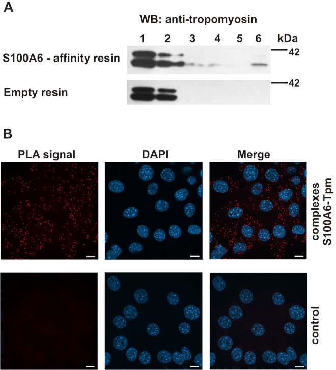
Figure 5.
Interaction of S100A6 with tropomyosin in NIH3T3 fibroblasts. (A) Pull-down assay with the use of protein lysate from NIH3T3 fibroblasts and with CNBr-Sepharose-S100A6 affinity resin (upper panel) or CNBr-Sepharose-empty resin (lower panel). Lanes: 1—input, 2—unbound fraction, 3—last wash, 4—first wash with 250 mM NaCl, 5—last wash with 250 mM NaCl, 6—elution. Fractions were analyzed by SDS-PAGE (10% gel) followed by immunoblotting developed with mouse monoclonal anti-tropomyosin antibody, Sigma Aldrich, clone TM311. (B) Presence of S100A6–Tpm complexes in NIH3T3 fibroblasts analyzed by PLA assay. Control represents the experiment in which ligase was omitted. Complexes of examined proteins are visualized in red; cell nuclei, stained with DAPI, are in blue. Scale bar is 10 μm.

