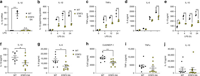
Fig. 5. STAT3 pS727 is required for LPS-induced cytokine expression.
a Peritoneal macrophages obtained from three mice per genotype were seeded at 5 × 10⁵ cells/well in and stimulated with LPS (100 ng/ml) for 24 h prior to analysis IL-1β mRNA expression. Data are presented as mean values ± SEM, ***p < 0.0001, n.s.—not significant, one-way ANOVA, Tukey’s multiple comparisons test. Peritoneal macrophages generated from individual WT and STAT3 mice (3 mice per genotype) were seeded at 1.5 × 10⁵ in triplicate. Macrophages were stimulated for 4, 12, and 24 h with 100 ng/ml of LPS and b cell lysates assayed for IL-1β and presented as IL-1β/mg of total protein **p = 0.0011, ***p < 0.0001, and cultured supernatants assayed for c TNF (4 h, ***p = 0.0007; 12 h **p = 0.0015; 24 h ***p = 0.0001), d IL-6 (***p < 0.0001), and e IL-10 (*p = 0.0168, ***p < 0.0001) by ELISA. Data presented are the mean of triplicate values from three individual mice (n = 3) per genotype per time point, Analyses 2 way ANOVA, Sidak’s multiple comparison tests. WT and STAT3 SA mice (n = 9 mice per genotype) were intraperitoneally treated with 10 mg/kg of LPS for 90 min and serum analysed for f IL-1β (*p = 0.0444), g IL-6 (*p = 0.0313), h MCP-1 (**p = 0.0046), i TNF (p = 0.3994) and j IL-10 (p = 0.8204) protein expression by ELISA or cytometric bead array. Data are presented as mean values ± SEM, Students unpaired t test, two-tailed.

