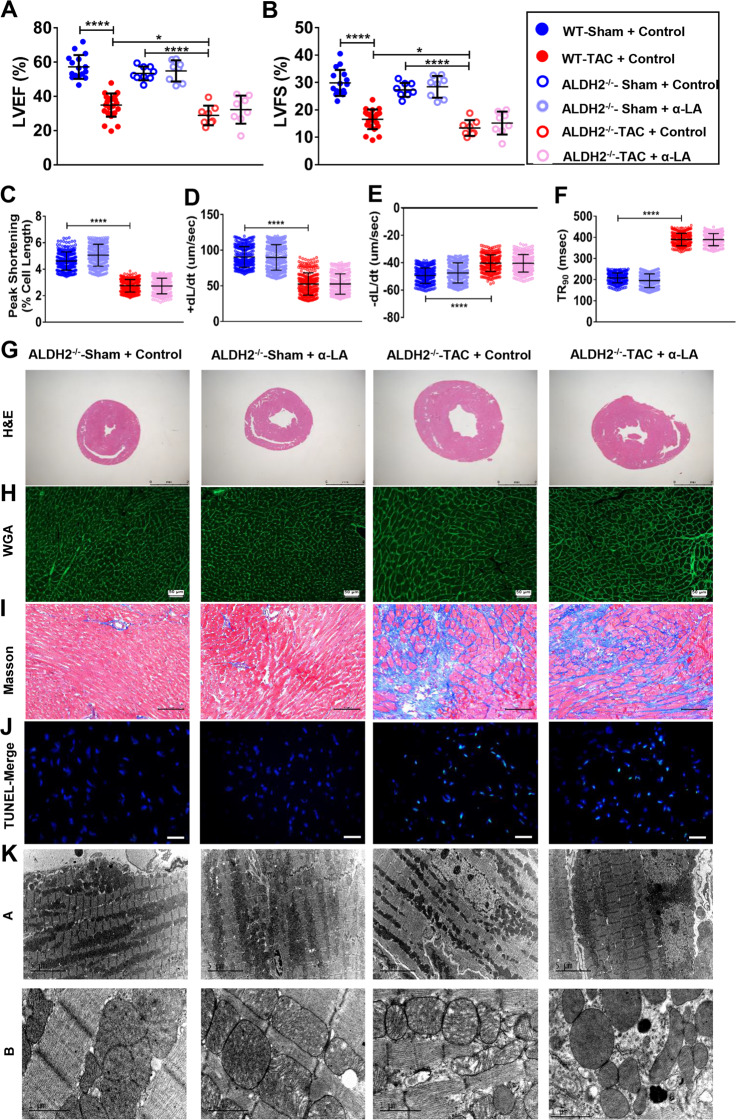
Fig. 5. α-LA treatment improves cardiac function in TAC mice via ALDH2-dependent activation of FUNDC1.
a Left ventricular ejection fraction (LVEF, %) (n = 8–20 mice per group); b left ventricular fractional shortening (LVFS, %) (n = 8–20 mice per group); c cardiomyocyte peak shortening (PS, normalized to resting cell length) (n = 300 cells per group); d maximal velocity of shortening (+dL/dt) (n = 300 cells per group); e maximal velocity of relengthening (−dL/dt) (n = 300 cells per group); f time-to-90% relengthening (TR90) in isolated cardiomyocytes (n = 300 cells per group); g hematoxylin–eosin (H&E, scale bar = 2 mm) staining; h wheat germ agglutinin (WGA, scale bar = 50 μm) staining; i Masson’s Trichome staining (scale bar = 100 μm); j terminal dexynucleotidyl transferase (TdT)-mediated dUTP nick end labeling staining (TUNEL, 400×, scale bar = 20 μm); k transmission electron microscopy (TEM) images of left ventricular, Up: scale bar = 5 μm, Down: scale bar = 1 μm. Mean ± SEM, *P < 0.05, ****P < 0.0001. Statistical analysis was carried out by a one-way ANOVA analysis followed by Tukey’s test for post hoc analysis.

