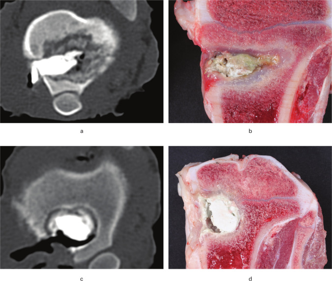
Fig. 1.
CT scans (cranio-caudal direction) and macroscopic images obtained ten and 12 days after revision surgery in a porcine model of Staphylococcus aureus osteomyelitis. Pigs were treated with either limited debridement (A + B) or extensive debridement (C + D) followed by injection of CERAMENT|G into the bone voids. a) Osteomyelitis with osteolysis and irregular borders of the lesion. b) CERAMENT|G surrounded by pus and fibrosis. c) Regular sclerotic border of the bone void. d) CERAMENT|G surrounded by a rim of fibrosis.

