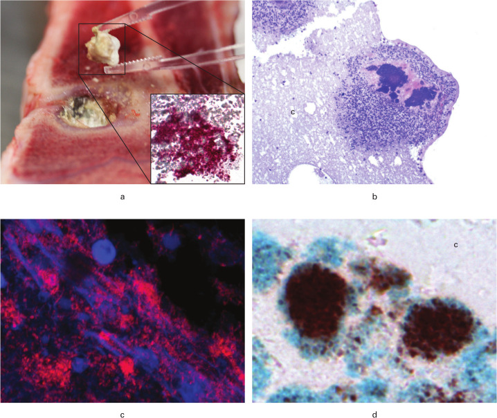
Fig. 3.
Visualization of bacteria ten and 12 days after revision surgery in a porcine model of Staphylococcus aureus osteomyelitis. Pigs were treated with either limited (A + C) or extensive (B + D) debridement followed by injection of CERAMENT|G into the bone voids. a) Macroscopic picture showing removal of CERAMENT|G. Insert: Immunohistochemistry (IHC) towards S. aureus of the CERAMENT|G removed in Figure 3a; red bacteria are demonstrated here (× 200). b) Bone void border showing bacterial aggregates surrounded by pink matrix and neutrophil granulocytes inside CERAMENT|G; haematoxylin and eosin (HE) stain (× 100). c) Peptide nucleic acid fluorescence in situ hybridization (PNA FISH) of CERAMENT|G showing red aggregates of S. aureus (× 630). d) Biofilm forming S. aureus in CERAMENT|G; red bacteria surrounded by a blue matrix is seen here, image produced with IHC combined with Alcian blue pH3 (× 400). c, CERAMENT|G.

