. 2020 Jul 28;61(8):731–735. doi: 10.3349/ymj.2020.61.8.731

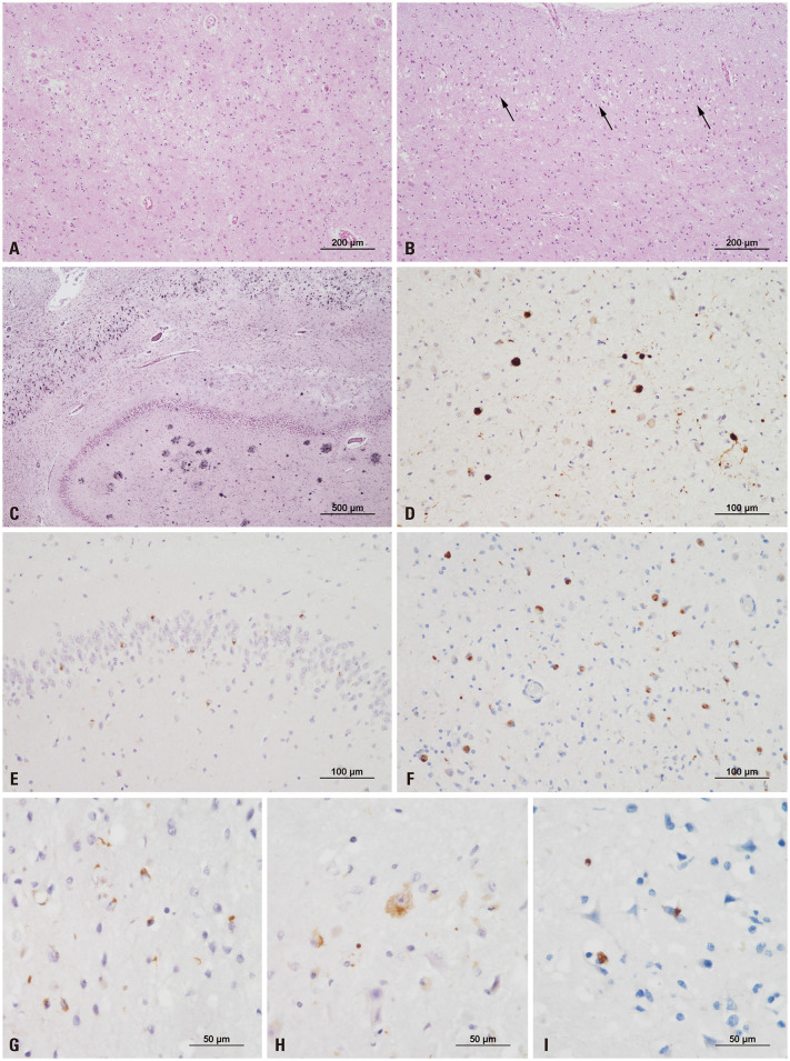
Fig. 3. Histopathological features of a case with LATE-NC. (A) The amygdala shows remarkable neuronal loss and gliosis. (B) Superficial microvacuolation in cortical layer II (black arrows) is observed in the inferior temporal cortex (A and B, hematoxylin and eosin; original magnification, ×100). (C) The hippocampus shows an abundance of neurofibrillary tangles and neuritic plaques, consistent with high Alzheimer disease neuropathologic changes (Gallyas silver stain; original magnification, ×40). (D) Alpha-synuclein immunostaining highlights abundant Lewy bodies solely in the amygdala, consistent with amygdala-predominant Lewy body disease. (E) Immunohistochemistry for TDP-43 indicates neuronal cytoplasmic inclusions (NCIs) in the dentate gyrus of the hippocampus. (F–H) Abundant NCIs and a few dystrophic neurites are observed in the amygdala (F), entorhinal cortex (G), and hippocampus (H). (I) NCIs are sparsely present in the insula (D–F, original magnification, ×200; G–I, original magnification, ×400).

Fig. 3