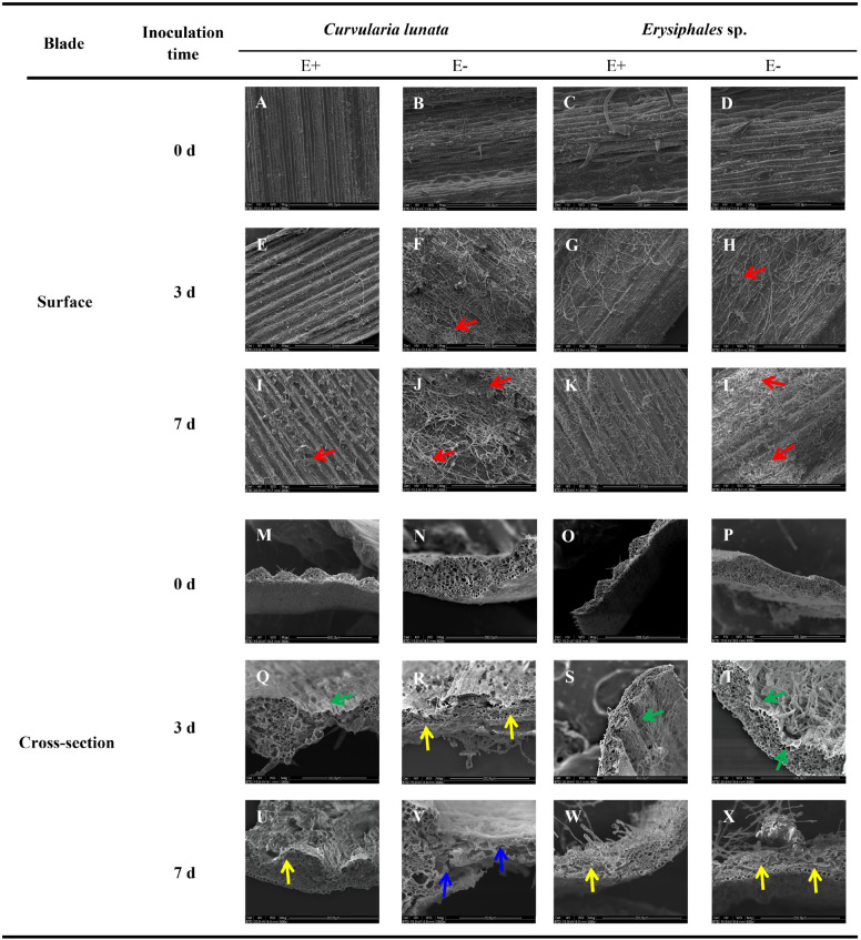
FIGURE 1.
Scanning electron microscopy observation of structures of E+ and E− A. sibiricum leaves infected by pathogens. Images A-L indicate the surface of E+ and E- leaves inoculated by C. lunata or Erysiphales sp. on Day 0 (A–D), 3 (E–H), and 7 (I–L), respectively. Images M-X indicate the cross-section of E+ and E- leaves inoculated by C. lunata or Erysiphales sp. on Day 0 (M–P), 3 (Q–T) and 7 (U–X), respectively. Red arrows indicate infection cushions; green arrows indicate crumpling cell walls; yellow arrows indicate collapsed cell walls, and blue arrows indicate fragmented cell walls.

