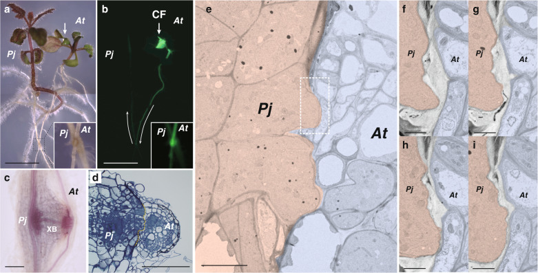
Fig. 1. Parasitism of P. japonicum.
a, b Parasitism between the roots of P. japonicum and Arabidopsis. The P. japonicum root parasitized the Arabidopsis root (insets). Transport of a symplasmic tracer dye, carboxyfluorescein (CF, colored in green), showed establishment of a symplasmic connection between the plants; light micrograph (a) and fluorescence image (b). Arrows indicate the site where CF dye was applied and the direction of transport. c, d Site where P. japonicum parasitized the Arabidopsis root. c Phloroglucinol staining showing xylem bridge formation (XB). d Cross-section of the parasitization site. The P. japonicum tissue invaded the Arabidopsis root tissues. Dashed line indicates the interface of parasitism. e Transmission electron micrograph of the interface between P. japonicum (pink) and Arabidopsis (blue). Partial tissue adhesion was observed at the interface. The dashed rectangle indicates the area of (f–i). f–i Serial sections at the interface between P. japonicum and Arabidopsis cells. The cell wall was partially digested. PjP. japonicum, AtArabidopsis. Scale bars, 5 mm (a, b), 100 μm (c, d), 10 µm (e), and 2 µm (f–i).

