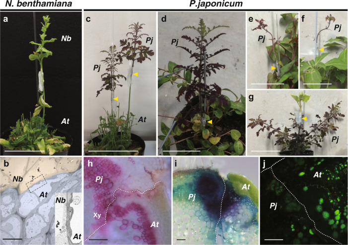
Fig. 2. Heterospecific grafting of N. benthamiana and P. japonicum.
a Grafting of N. benthamiana onto Arabidopsis at 28 days after grafting (DAG). b Transmission electron micrograph of the graft junction between N. benthamiana (pink) and Arabidopsis (blue). Inset represents a magnified image of the area of the dashed rectangle. c–g Grafting of P. japonicum with diverse plant species; grafts of P. japonicum as the scion onto stems of Arabidopsis at 28 DAG (c), Vinca major at 52 DAG (d), Houttuynia cordata at 30 DAG (e), and Pachysandra terminalis at 30 DAG (f), and graft of Cucumis sativus as the scion onto P. japonicum stock at 30 DAG (g). Arrowheads indicate grafting points. h Cross-section of the graft junction of P. japonicum/Arabidopsis. Phloroglucinol staining showing xylem formation in the graft region (Xy). i Cross-section of the graft junction of P. japonicum/Arabidopsis stained with toluidine blue applied to the Arabidopsis stock at 14 DAG. Detection of dye transport from Arabidopsis to P. japonicum demonstrated establishment of apoplastic transport. j Symplasmic transport establishment was confirmed using carboxyfluorescein (CF). CF was applied in the diacetate form to the leaves of the Arabidopsis stock and a cross-section of the graft junction of P. japonicum/Arabidopsis was observed. The CF fluorescence was detected in P. japonicum tissues. PjP. japonicum, AtArabidopsis. Scale bars, 5 cm (a, c–g), 1 µm (b), and 100 μm (h–j).

