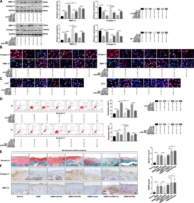
Fig. 5. CircRNF121/miR-665/MYD88 axis mediates ECM degradation, proliferation, and apoptosis in OA progression.
a MMP-13 and collagen II were detected by western blot. b Expression of MMP-13 and collagen II was presented by immunofluorescence staining. c The ki-67 intensity was shown by immunofluorescence staining. d The apoptotic rates of treated chondrocytes were measured by flow cytometry. Viable and nonviable apoptotic cells are regarded as apoptotic cells. e Histologic section of cartilage structures of SD rats was stained by safranin-O in each group. f The MMP-13 and collagen II levels in vivo were measured by IHC. Data were means ± SD of three independent assays (*P < 0.05).

