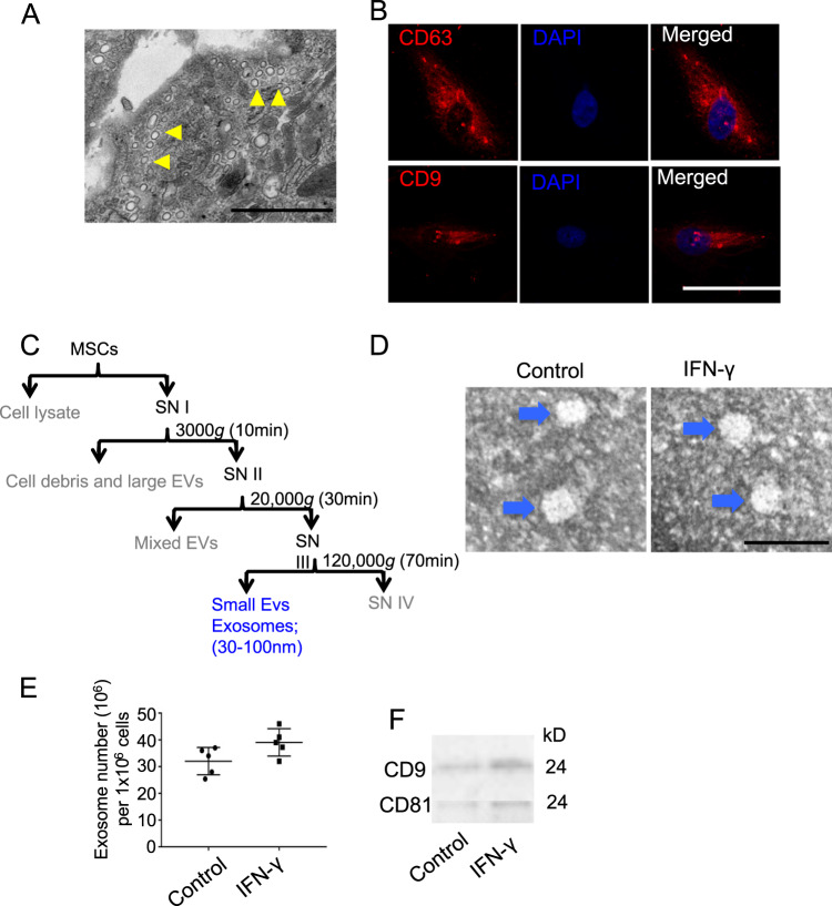
Fig. 1. Characteristic of exosomes isolated from mouse BMMSCs.
a Microvesicles in MSCs were analyzed by TEM. b Immunofluorescent staining of CD63 and CD9 in MSCs. c Differential centrifugation procedure for the isolation of exosomes from culture supernatants of MSCs (SN). d Isolated exosomes from control and IFN-γ-primed MSCs were analyzed by TEM. e Exosomes numbers isolated from control and IFN-γ-primed MSCs was analyzed by EXOCEP exosome quantitation kit. f The expression of CD81 and CD9 in exosomes from control and IFN-γ-primed MSCs were analyzed by western blotting. Scale bar a: 500 nm, b: 20 μm, and d: 100 nm. *P < 0.05, **P < 0.01, ***P < 0.001.

