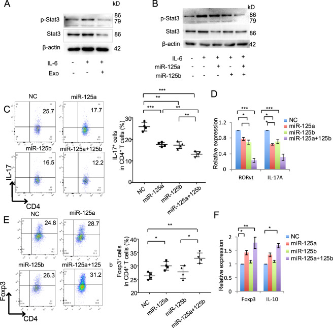
Fig. 4. MiR-125a and miR-125b inhibited Th17 cell differentiation.
a The expression of p-Stat3 and Stat3 in T cells after IL-6 stimulation with or without 50 ng/ml exosomes treatment, as assessed by western blot. b The expression of p-Stat3 and Stat3 in T cells with the treatment as indicated. c Th17 cell differentiation treated by miR-125a (20 nmol), miR-125b (20 nmol), or miR-125a and miR-125b combination mimics for 3–4 days, analyzed by flow cytometry. d The expression of RORγt and IL-17 in Th17 cells in control, miR-125a, miR-125b, or miR-125a and miR-125b combination mimics treatment groups, analyzed by qPCR. e Treg cell differentiation treated by miR-125a (20 nM), miR-125b (20 nM), or miR-125a and miR-125b combination mimics, as assessed by flow cytomotry. f The expression of Foxp3 and IL-10 in in control, miR-125a, miR-125b, or miR-125a and miR-125b combination mimics treatment groups, as assessed by qPCR. Exo exosomes, NC negative control miRNA. *P < 0.05, **P < 0.01, ***P < 0.001.

