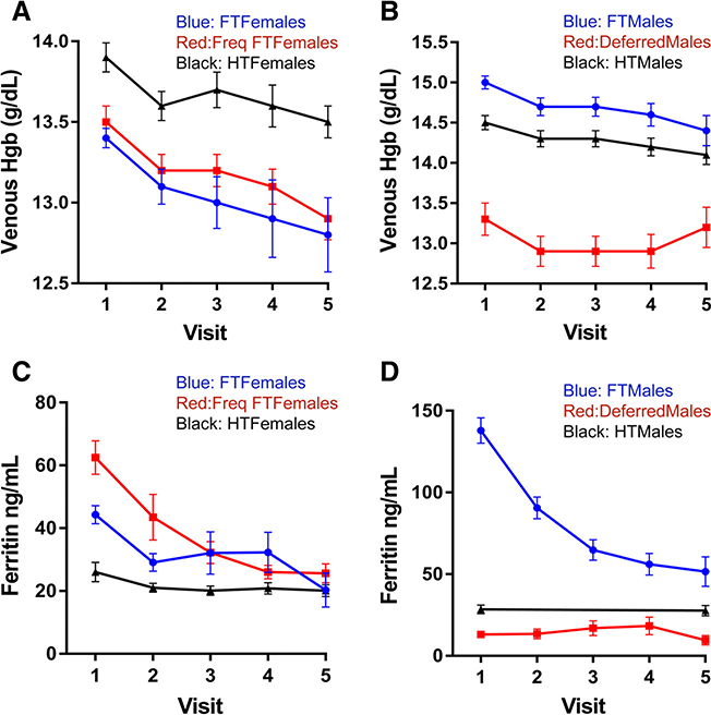
Fig. 2.
Change in hemoglobin and ferritin over multiple donations in different analysis groups. (A) Venous hemoglobin in female groups; (B) venous hemoglobin in male groups; (C) ferritin in female groups; and (D) ferritin in male groups. In A and C, blue lines are first- time/reactivated (FT) females; red lines are frequent first-time (Freq FT) females; and black lines are high-intensity (HT) females. In B and D, blue lines are first-time/reactivated (FT) males; red lines are deferred males; and black lines high-intensity (HT) males error bars represent standard error of the mean. [Color figure can be viewed at wileyonlinelibrary.com]

