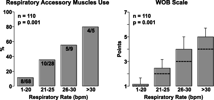
Fig. 2.
Left graph shows the percentage of patients who had activation of at least one of the accessory muscles assessed by the work breathing scale as a function of respiratory rate. Right graph shows the mean and standard deviation of the work breathing scale as a function of respiratory rate with the discontinuous line indicating the contribution of the respiratory rate alone (right). Analysis performed in 110 patients. Overall differences were analyzed using SigmaPlot 12.5 by chi-square on the left and by one-way analysis of variance on the right

