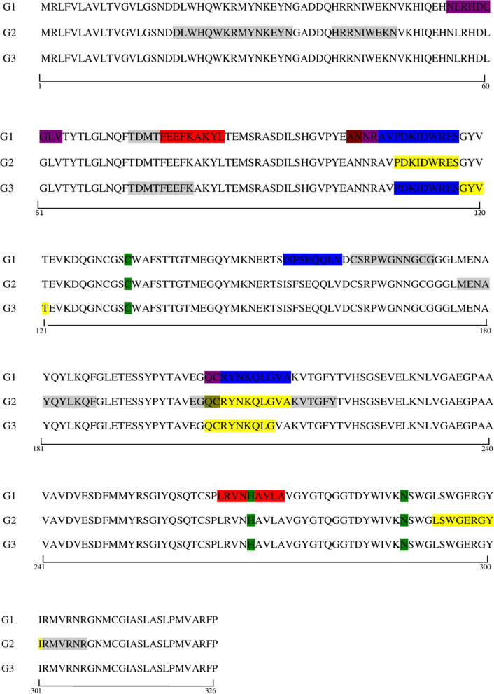
Fig. 5.
Localization of the peptides recognised in the linear FhCL1 sequence and comparison between groups and time points. Key: green, active sites of the protein; red, epitopes binding after vaccination and before infection (G1: peptide numbers 39, 133); blue, epitopes binding at the early stage of the infection (G1: peptide numbers 54-55-77-103); yellow, epitopes binding at the late stage of the infection (G2: peptide numbers 55-103-147; G3: peptide numbers 56-57-102); olive green, epitopes at early and late stage of the infection (G2: peptide number 102); purple, epitopes binding after vaccination and at the early infection (G1: peptide numbers 28-53-102); brown, epitopes binding at all time points (G1: peptide number 52); grey, non-specific binding of epitopes

