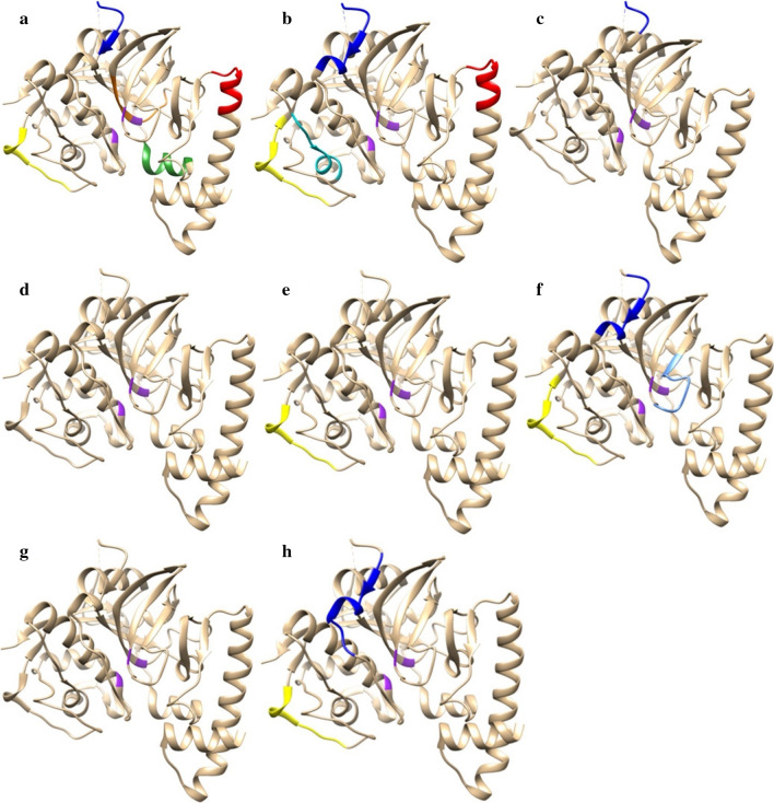
Fig. 6.
Epitope recognition and localisation in the 3D FhCL1 structure. a G1 (4 wav): red (aa 55–63, NLRHDLGLV); forest green (aa 77–84, FEEFKAKYL); blue (aa 102–112, ANNRAVPDKID); yellow (aa 203–211, QCRYNKQLG); orange (aa 265–273, LRVNHAVLA). b G1 (4 wai, early stage): red (aa 55–63, NLRHDLGLV); blue (aa 102–116, ANNRAVPDKIDWRES); cyan (aa 153–161, ISFSEQQLV); yellow (aa 203–213, QCRYNKQLGVA). c G1 (12 wai, late stage): blue (aa 102–110, ANNRAVPDK). d G2 (4 wav): no peptides were recognised at this time point. e G2 (4 wai, early stage): yellow (aa 203–211, QCRYNKQLG). f G2 (12 wai, late stage): blue (aa 108–116, PDKIDWRES); yellow (203–213, QCRYNKQLGVA); cornflower blue (293–301, LSWGERGYI). g G3 (4 wai, early stage): no peptides were recognised at this time point. h G3 (12 wai, late stage): blue (aa 110–120, KIDWRESGYVT); yellow (aa 203–211, QCRYNKQLG). Purple: active sites of the protein

