
Figure 1.
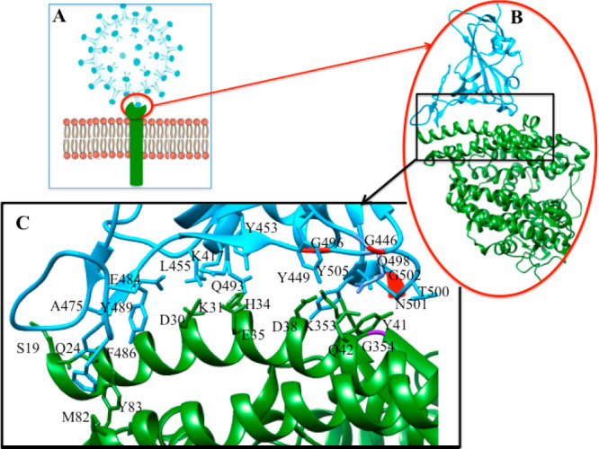
(A) Cartoon represents the attachment of SARS-CoV-2 (sky blue sphere with spike) and hACE2 (green rod). (B) Crystal structure of spike protein (sky blue) bound hACE2 (green) complex (PDB: 6m0j),2b (C) Selective amino acid residues at the contact interface between spike protein and hACE2 are highlighted with numbering (for simplicity, noncovalent interactions lines are omitted). H-bonding interaction: K417(COVID-19)---D30(ACE2), G446(COVID-19)---Q42(ACE2), Y449(COVID-19)---D38(ACE2), Y449(COVID-19)---Q42(ACE2), Y453(COVID-19)---H34(ACE2), A475(COVID-19)---S19(ACE2), F486(COVID-19)---Y83(ACE2), N487(COVID-19)---Q24(ACE2), Q493(COVID-19)---E35(ACE2), G496(COVID-19)---D38(ACE2), G496(COVID-19)---K353(ACE2), Q498(COVID-19)---Q42(ACE2), T500(COVID-19)---Y41(ACE2), N501(COVID-19)---Y41(ACE2). Electrostatic interaction: G502(COVID-19)---K353(ACE2), K417(COVID-19)---D30(ACE2), E484(COVID-19)---K31(ACE2), L455(COVID-19)---H34(ACE2). Hydrophobic interaction: F486(COVID-19)---M82(ACE2), Y489(COVID-19)---K31(ACE2), Y505(COVID-19)----K353(ACE2).