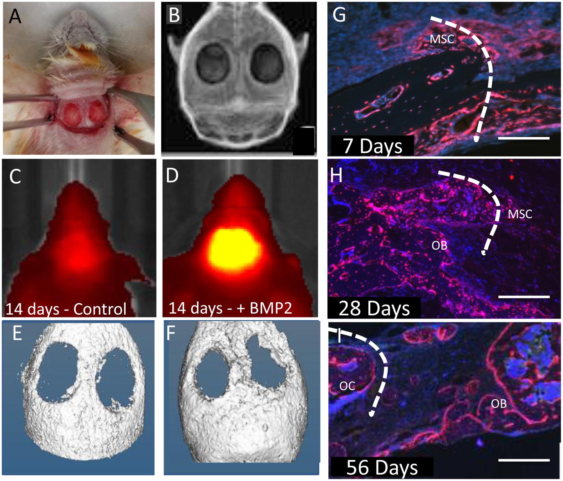
Figure 2: Osterix-Cherry expression provides a read out of putative bone formation before mineralization and correlating with neo-osteogenesis.

(A) Osterix mCherry mouse prepared for insertion of either collagen sponge or nanocomposite hydrogel. (B) X-ray of bilateral calvarial defect post surgery. (C) Osterix-mCherry mouse IVIS image 14 days post surgery with only collagen sponge inserted. (E) Corresponding μCT image. (D) Osterix-mCherry mouse IVIS image 14 days post surgery with collagen sponge infused with rhBMP-2. (F) Corresponding μCT image. (G) Calvarial defect treated with a collagen sponge infused with rhBMP-2 7 days post surgery showing mesenchymal stem cells (MSC) infiltrating the defect site (scale bar = 500μm). (H) Calvarial defect treated with a collagen sponge infused with rhBMP-2 28 days post surgery showing MSCs and Osterix-mCherry Osteoblasts (OB) (scale bar = 500 μm). (I) Calvarial defect treated with a collagen sponge infused with rhBMP2 56 days post surgery showing Osterix-mCherry Osteoblasts (OB) and Osteocytes (OC) (scale bar = 500 μm). Dotted line represents defect edge.
