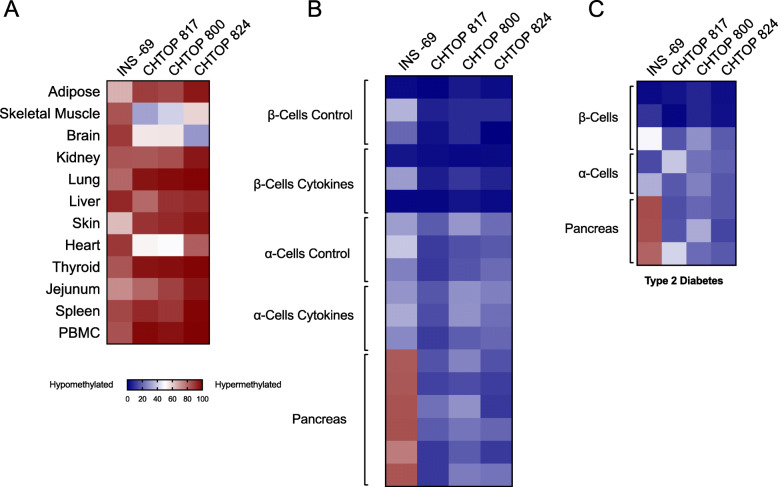
Fig. 4.
Relative abundance of differentially methylated CHTOP and INS DNA in human tissue samples by droplet dPCR. DNA from the indicated human tissues was isolated, bisulfite treated, and differentially methylated CHTOP-817, − 800, − 824, and INS DNA levels were quantitated by droplet dPCR. Data are displayed as a heatmap (blue = unmethylated, red = methylated). a Non-pancreatic tissues, each from a single donor. b Flow-sorted β cells (N = 3 donors) treated with and without proinflammatory cytokines (IL-1β and IFN-γ), α cells (N = 3 donors) treated with and without proinflammatory cytokines, and total pancreas (N = 6 donors). c Flow-sorted β cells (N = 3 donors) treated with and without proinflammatory cytokines, α cells (N = 2 donors) treated with and without proinflammatory cytokines, and total pancreas (N = 3 donors) from subjects with type 2 diabetes

