Fig. 4.

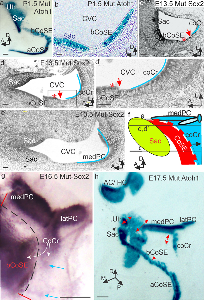
Mutant cochlear and crista sensory epithelia make contact. (a, b) P1.5 Whole mount (a) and section (b) stained for Atoh1 expression in hair cells. Note the crista is parted from the basal cochlear sensory epithelium (bCoSE) at this age and is not included. Black line in (a) marks the plane of section in b where the basal CoSE approaches the saccular macula (Sac) and joins it in an adjacent section (not shown). (c, d, d’,e) Selected horizontal serial sections cut from an E13.5 ear stained for Sox2 expression in planes indicated in f. Blue lines approximate crista sensory epithelium. Red asterisks mark the abneural edge of the basal cochlear sensory epithelium. Red arrows mark the region of contact between crista and cochlear sensory epithelia. The cochlear sensory epithelium is artifactually broken in (d). (d’) shows the boxed area in (d). In (e), the plane of section is dorsal to the cochlear sensory epithelium. (f) Diagram illustrating the association of the crista sensory epithelium with the basal cochlear sensory epithelium along the dashed red line. Here the cochlear sensory epithelium will lack an outer hair cell band and cochlear Bmp4 expression will be patchy. (g) Whole mount of an E16.5 posterior and cochlear crista stained for Sox2 expression. White arrows mark the proposed directions of crista propagation by lateral induction. Blue arrows mark distal patches of Sox2 expression. Note the broad and elongated medial arm of the posterior crista (medPC). Dotted black line marks the now separating zone of contact with the basal cochlear epithelium. (h) By E17.5, the cochlear crista and medial posterior cristae are separated from the basal cochlear sensory epithelium, but the former contact zone can still be recognized (double headed red arrows). Abbreviations: aCoSE, apical cochlear sensory epithelium; AC/HC, anterior and horizontal cristae; bCoSE, basal cochlear sensory epithelium; coCr, cochlear crista; CVC, common vestibular cavity; latPC, lateral arm of the posterior crista; medPC, medial arm of the posterior crista; Sac, saccular macula; Utr, utricle. Bars in a, g, and h are 100 μm. All others are 10 μm
