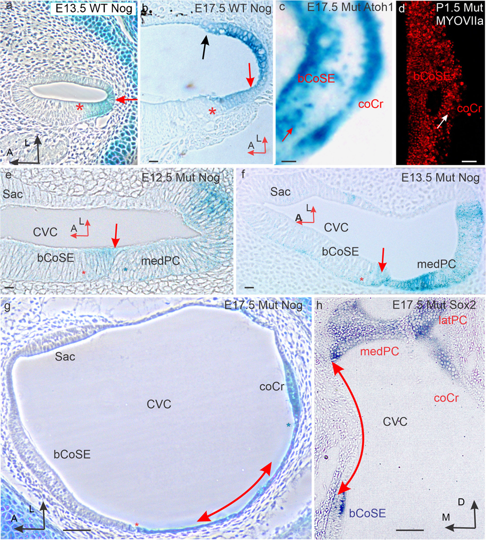
Fig. 5.

A Nog-expressing epithelium separates basal cochlear sensory epithelium from crista sensory epithelium. (a) Section across the cochlear duct of an E13.5 wild-type ear stained for Nog expression (red arrow). Red asterisk marks the site of the cochlear sensory epithelium. (b) Similar section across an E17.5 wild-type duct stained for Nog expression in the outer spiral sulcus. Red asterisk marks the site of the cochlear sensory epithelium. A black arrow marks the edge of the stria vascularis identified by black melanocytes in its forming intermediate layer. A red arrow marks the outer spiral sulcus. (c) Atoh1 stained cochlear duct from the E17.5 mutant ear in Fig. 2a’. Note several hair cells (e.g., red arrow) are located in the epithelium separating the cochlear sensory epithelium from the crista sensory epithelium. (d) Flat mount of the basal CoSE of a P1.5 mutant immunostained for MYOSIN VIIa to label maturing hair cells. White arrow marks the abneural margin of the basal cochlear sensory epithelium. Note scattered hair cells to the right of the margin. (e) Horizontal section across the posterior crista and basal cochlear sensory epithelium of an E12.5 mutant ear. The medial arm of the crista (blue asterisk) and cochlear sensory epithelium (red asterisk) are in direct planar contact (red arrow). Nog expression stains the interface between the two. (f) similar section from an E13.5 ear. The contacting edge of the crista is thinning. (g) Horizontal section across the common vestibular cavity (CVC) of an E17.5 mutant ear stained for Nog expression. A red asterisk marks the abneural (lateral) margin of the basal cochlear sensory epithelium, and a blue asterisk marks the facing margin of the cochlear crista. The double headed arrow parallels the Nog stained epithelium separating the cochlear sensory epithelium from the crista sensory epithelium. (h) Vertical section across the common vestibular cavity of an E17.5 mutant ear stained for Sox2 expression. The double-headed red arrow parallels the unstained non-sensory epithelium separating the cochlear sensory epithelium from the crista. Abbreviations: bCoSE, basal cochlear sensory epithelium; coCr, cochlear crista; CVC, common vestibular cavity; latPC, lateral arm of the posterior crista; medPC, medial arm of the posterior crista; Sac, saccular macula. Bars in c, d are 100 μm. All others are 10 μm
