Fig. 6.
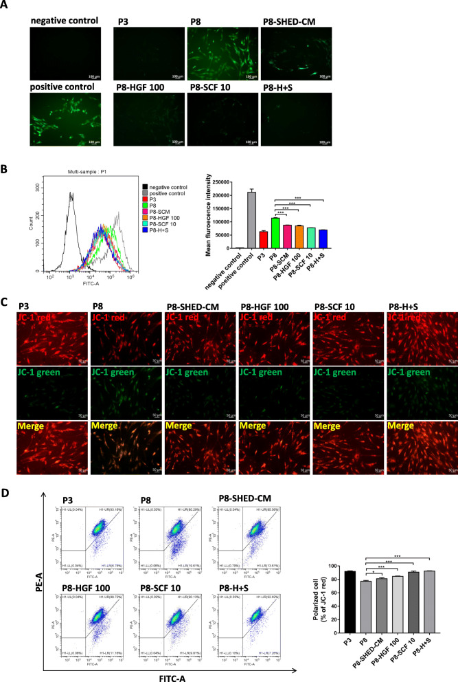
HGF and SCF reduced ROS accumulation and preserved mitochondrial function of hBMSCs during long-term expansion. a Representative images of ROS levels in negative control, positive control (stimulated with 1 mM H2O2 for 1 h), passage 3 (P3), and passage 8 (P8) hBMSCs cultured in SHED-CM (P8-SHED-CM) and treated with 100 ng/ml HGF (P8-HGF 100), 10 ng/ml SCF (P8-SCF 10), and the combination of 100 ng/ml HGF and 10 ng/ml SCF (P8-H+S). b Flow cytometry results of ROS levels and mean fluorescent intensity in hBMSCs per treatment group. c Representative images of mitochondrial membrane potential in hBMSCs per treatment group detected by JC-1 probe. The J-aggregates produced red fluorescence (JC-1 red); the monomer produced green fluorescence (JC-1 green). d Flow cytometry results of mitochondrial membrane potential and corresponding percentage (%) of polarized cell per treatment group detected by JC-1 probe. *P < 0.05, ***P < 0.001

