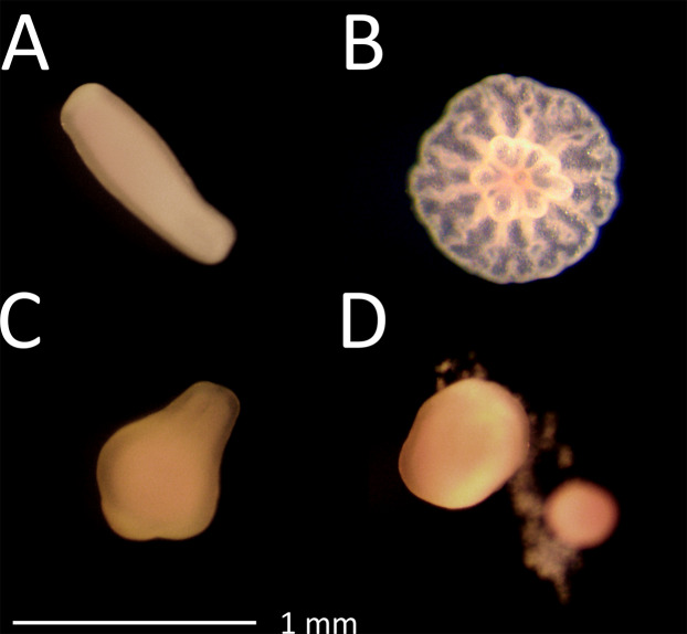
Figure 1. Photographs after 48 h exposure.
(A) Planula larvae in control treatment; (B) attached post-metamorphosis polyp in control treatment; (C) larvae exposed to 228 µg L−1 propiconazole showing slightly abnormal shape but still moving and (D) larvae exposed to 56.3 µg L−1 chlorothalonil showing rupturing of cells.

