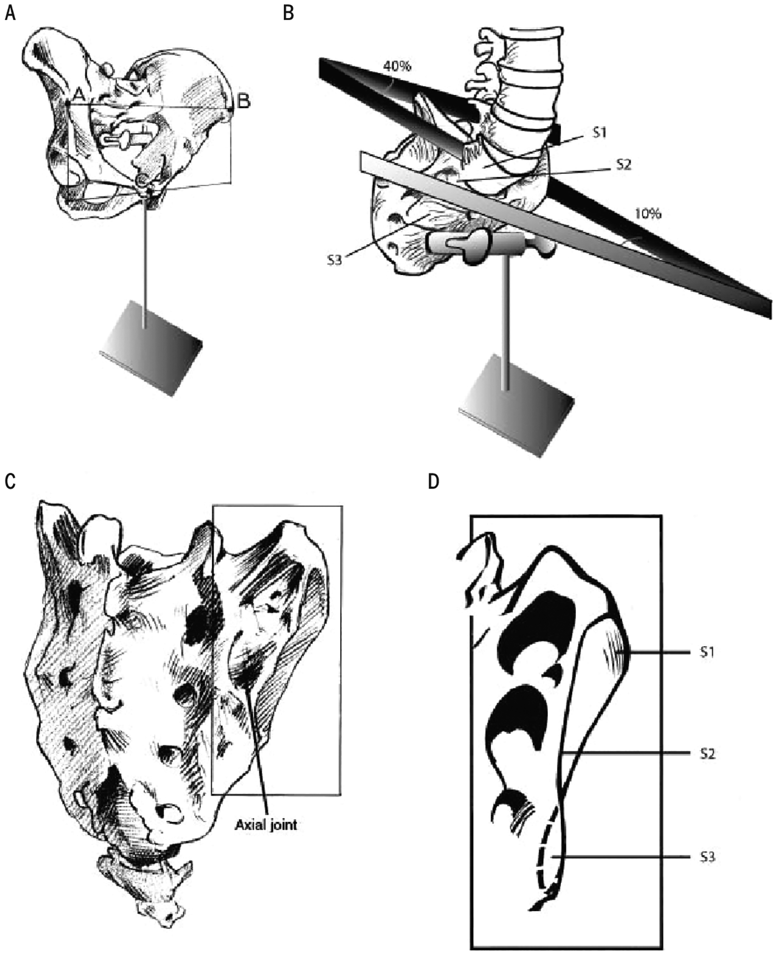
FIGURE 5.

(A) The pelvis in erect posture. (B) View of the sacrum from the ventrolateral side, showing the different angles between left and right sacral articular surfaces. (C) Dorsolateral view of the sacrum. The pointer indicates a cavity in the sacrum, in which an iliac tubercle fits, called the “axial” sacroiliac joint. (D) Sacral articular surface at the right side. The different angles reflect the propeller-like shape of an adult sacroiliac joint.
