
Figure 2.
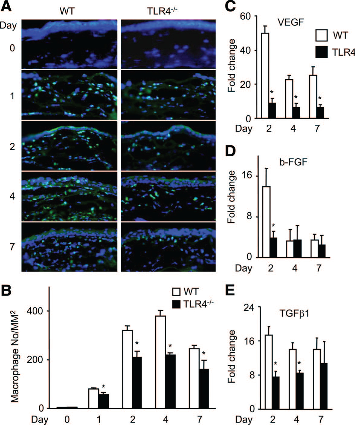
Macrophage accumulation and angiogenic factor expression in injured corneas. A, Corneal tissues from WT mice (left panels) or TLR4−/− (right panels) mice were stained with fluorescein isothiocyanate–conjugated anti-F4/80 monoclonal Abs. Magnification ×400. B, The numbers of infiltrating F4/80-positive macrophages were determined, and the means±SEM are shown (n=6). *P<0.05 vs WT mice. C to E, Angiogenic factor expression in injured corneas. The mRNA expression of VEGF (C), b-FGF (D), and TGFβ1 (E) in wound sites was determined by quantitative reverse transcription–PCR. Results are expressed as mean±SEM of fold increase over control. *P<0.05 vs WT mice.