
Figure 3.
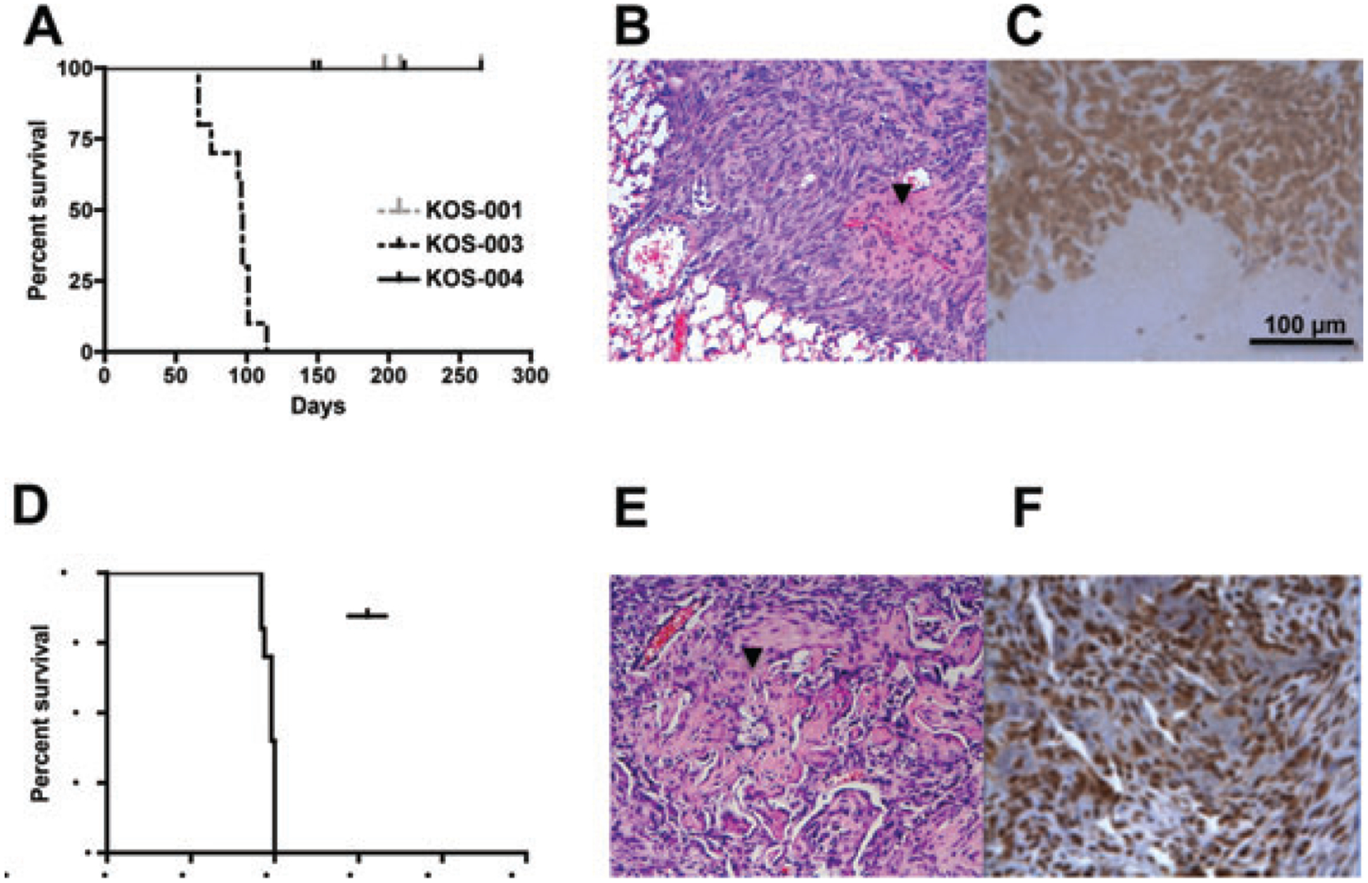
Assessment of in vivo metastatic phenotype of canine osteosarcoma (OS) cell lines. Overall survival of canine OS cells in xenograft metastasis model. (A) Kaplan–Meier survival curves demonstrate progression to spontaneous metastasis in KOS-003 cells following orthotopic delivery of tumour cells to mice. No spontaneous pulmonary metastases were seen in KOS-001 and KOS-004 over 1-year observation period. (B) H&E staining of the resultant metastases were consistent with OS. (C) Spontaneous lung metastasis tumours showed positive ALP staining. (D) The metastatic biology of the KOS-003 was confirmed using experimental metastasis via tail vein injection. (E) H&E staining of experimental metastasis again show osteoid production (arrow head) and (F) ALP staining. Scale bar = 100 μm.