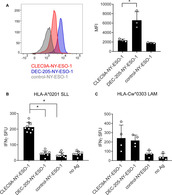
Figure 2.
Cross-presentation of NY-ESO-1 epitopes following uptake of CLEC9A-NY-ESO-1 by CD141+ DC. (A) Binding of chimeric Abs to in vitro cultured CD141+ DC shown as representative histograms (left) and the median fluorescence intensity (MFI) collated from three independent donors (right); *p=0.04. (B) Cross-presentation of the HLA-A*0201-restricted NY-ESO-1 SLL epitope following uptake of CLEC9A-NY-ESO-1, DEC-205-NY-ESO-1, control-NY-ESO-1 chimeric Abs or no Ag by in vitro cultured HLA-A*0201+ CD141+ DC. Cross-presentation was measured by interferon γ (IFNγ) production by NY-ESO-1 SLL-specific CD8+ T cells by ELISPOT (spot forming units (SFU) per 10,000 T cells). (C) Cross-presentation of the HLA-Cw3-restricted NY-ESO-1 LAM epitope following uptake of chimeric Ab. Cross-presentation was measured by IFNγ production by NY-ESO-1 LAM-specific CD8+ T cells by ELISPOT (SFU per 10,000 T cells). Data shown are the mean±SD collated from replicates of four independent donors. *p<0.0001 by one-way analysis of variance and Tukey’s multiple comparison test. Ab, antibody; Ag, antigen; DC, dendritic cell; NY-ESO-1, New York esophageal squamous cell carcinoma 1.

