Figure 6.
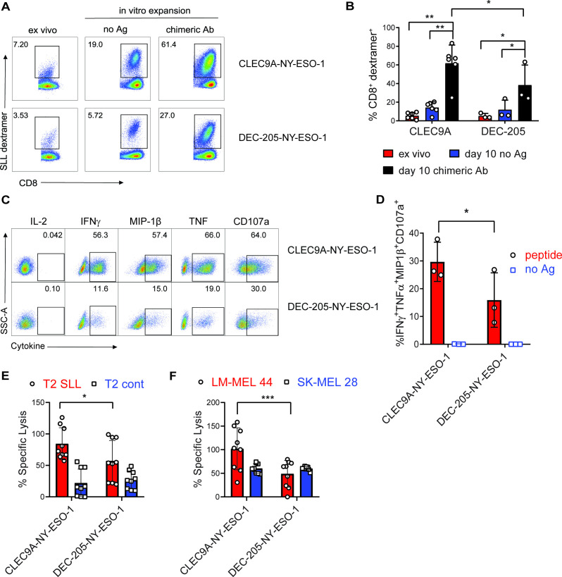
CLEC9A-NYESO-1 primes naïve NY-ESO-1-specific CD8+ T cells for effector function. Humanized mice were vaccinated with CLEC9A-NY-ESO-1 or DEC-205-NYESO-1 in the presence of poly I:C. (A) Percentage of NY-ESO-1 SLL dextramer+ CD8+ T cells in splenocytes 1 week after vaccination (ex vivo) and again after 10 days in vitro expansion in the presence of no Ag or the same chimeric Ab used for immunization shown as representative plots. (B) Mean percentage of NY-ESO-1 SLL-specific CD8+ T cells±SD from individual mice. (C) Cytokine secretion, from CD8+ T cells that were primed and expanded with CLEC9A-NY-ESO-1 or DEC-205-NY-ESO-1, following a 6-hour restimulation with SLL peptide or no Ag as representative flow cytometry plots for each cytokine. (D) Boolean gating analysis of the percentage of CD8+ T cells producing all four effector molecules (IFNγ, MIP-1β, TNF and CD107a) as the compiled mean±SD from individual mice. (E) Lysis of T2 target cells pulsed with NY-ESO-1 SLL peptide or irrelevant control peptide (NLVPMVATV) pulsed T2. (F) Lysis of HLA-A*0201+ NY-ESO-1+ (LM-MEL-44) and HLA-A*0201-NY-ESO-1- (SK-MEL-28) melanoma cell lines by CD8+ T cells primed and expanded with CLEC9A-NY-ESO-1 or DEC-205-NYESO-1. Compiled replicate means±SD from groups of n=3 individual mice are shown. *p<0.05, **p<0.0001, ***p<0.0005 by two-way analysis of variance and Tukey’s multiple comparison test. Ab, antibody; Ag, antigen; IFNγ, interferon γ; NY-ESO-1, New York esophageal squamous cell carcinoma 1; poly I:C, polyinosinic:polycytidylic acid; TNF, tumor necrosis factor.

