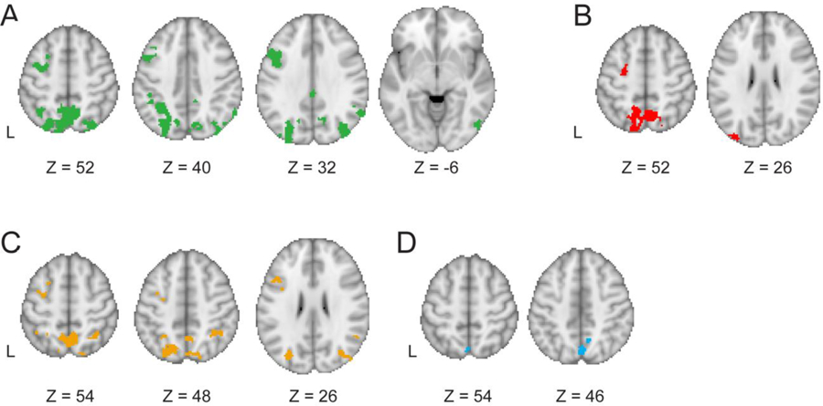
Figure 5.

Regions for which brain activity positively scaled with trial-by-trial (A) unsigned prediction errors. (B) When contrasting shift > hold trials with the PE regressor in the GLM, we found significant activity within mSPL, left FEF, and left superior lateral occipital cortex. Regions for which brain activity positively scaled with (C) signed shift trial prediction errors and (D) signed hold trial prediction errors.
