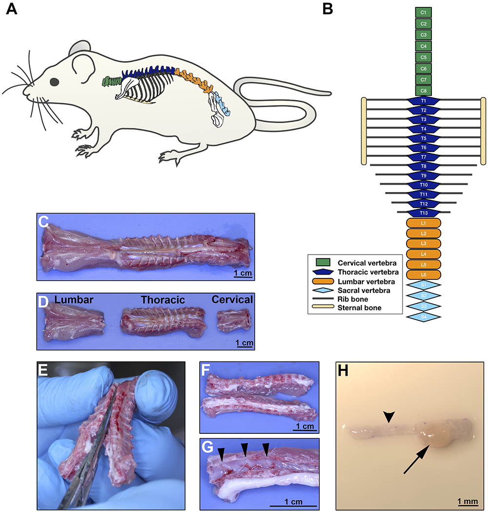
Figure 2: DRG extraction on day 8.

A. Rat schematic illustrating the anatomical location of the spine. B. Diagram of the rat vertebrae configuration showing different body regions; green for cervical, dark blue for thoracic, orange for lumbar and light blue for sacral vertebrae. C-D. Ventral aspect of the rat spine after surgical excision; separation of the regions as illustrated in B. E. Dissection of the vertebra to open the spinal cord canal, separating the vertebral body into two lateral sections containing the DRG. Section should cut through the dorsal and ventral aspect of each vertebral bone at the midline. F. Gross aspect of opened thoracic spine. G. After the spinal cord is displaced, DRG are easily visible in the vertebral canals (arrow heads pointing 3 DRGs). H. Stereomicroscopic image of one DRG (arrow) with the corresponding axon bundles (arrow head). Scale bars: C, D, F, and G, 1 cm; H, 1 mm.
