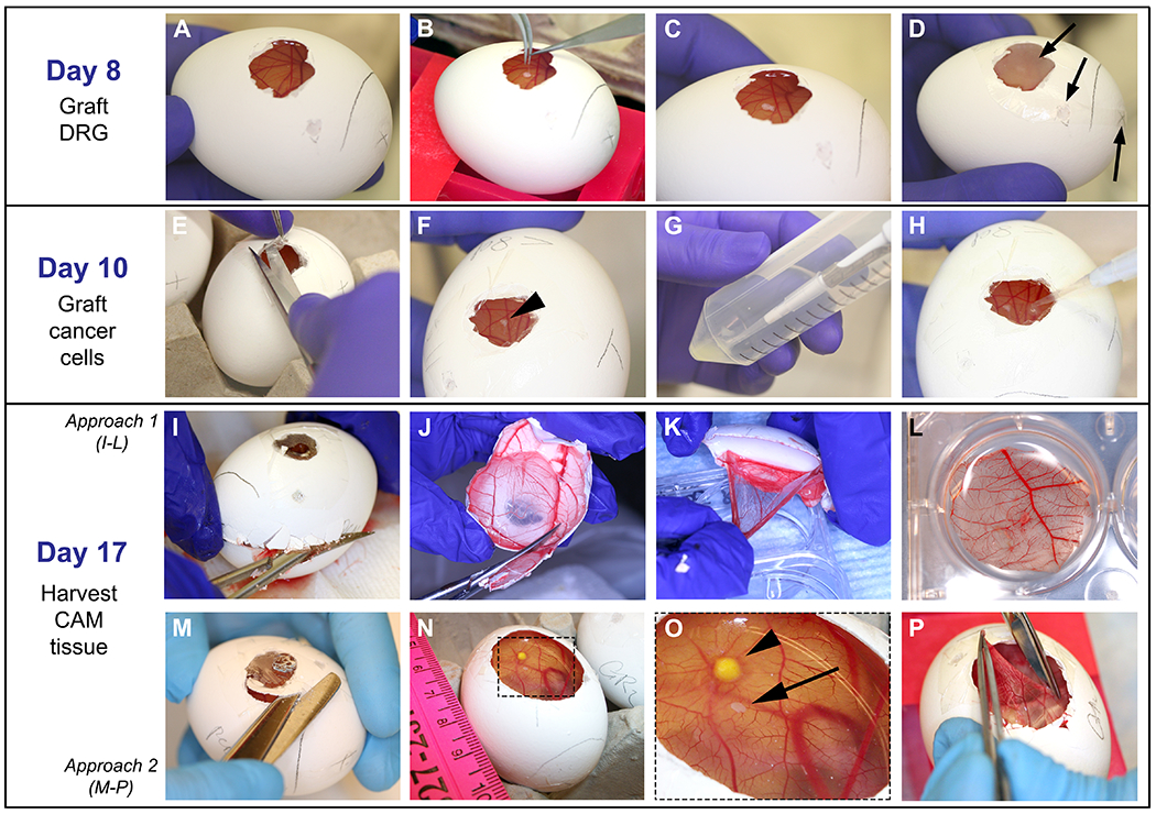
Figure 4: Grafting of DRG, cells and harvesting of CAM: On day 8:

A. CAM easily observed after paraffin wax membrane removal. B-C. With fine forceps, DRG is placed onto the CAM. D. Egg is covered with film dressing and put in the incubator; arrows point to the openings that are covered. On day 10: E-F. Film dressing is removed and DRG is located (arrow head on F). G-H. 5 μL of cell solution is dropped onto the CAM at a ~2 mm distance from the DRG. On day 17: I-L and M-P demonstrate two different approaches used to harvest the CAM. I. Egg shell is opened with a fine scissor starting on the air sac drilled perforation until the upper half of the egg is removed. J. Egg shell containing the CAM is reduced in size to approximately 3 cm. K-L. With fine forceps, CAM is detached from the egg shell and placed in PFA. M-O. Widening of the operating window is performed to visualize the DRG and cancer cells on the CAM. Arrowhead points to the tumor and arrow points to the DRG. P. The CAM is grasped with fine forceps, cut out with a sharp scissor, and placed in PFA as shown in L.
