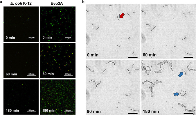
FIG 2.
Microscopy of persisters from E. coli K-12 and Evo3A during resuscitation after ampicillin treatment. (a) Cells exposed to ampicillin (∼15× MIC) for 5 h were resuspended in fresh LB medium and observed by epifluorescence microscopy at indicated time points during resuscitation. Green cells are live cells while red cells are dead cells (defined as those with compromised membrane). (b) Time-lapse microscopy of Evo3A during resuscitation on LB pads. Figures are representative phase-contrast images of recovering ampicillin-treated Evo3A cells on LB agar pads, and the panel at t = 180 min shows the incipient colony formation from a filament. Red arrow shows a lysed filament, and blue arrows indicate cell division events that are slower than the other cells. Bars, 50 μm.

