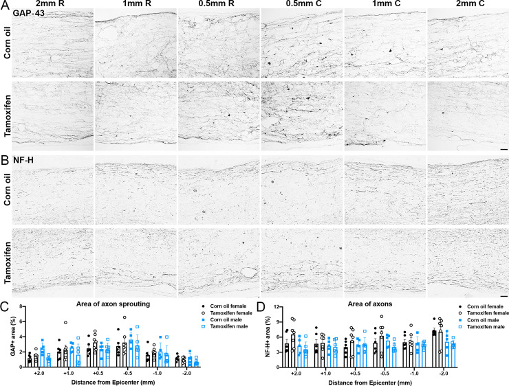
Fig 5. Delayed tamoxifen treatment does not alter total axon area after SCI.
(A-B) Representative images of longitudinal spinal cord sections immunolabeled for (A) GAP-43 and (B) neurofilament-heavy (NF-H). Scale bar = 20 μm. (C-D) Percent area of (C) GAP-43 and (D) NF-H+ axons in the lateral white matter 2 mm, 1 mm, and 0.5 mm rostral and caudal to the lesion epicenter. No significant differences are reported between groups. Data presented as mean + SEM. Two-way repeated measures ANOVA with Šídák post hoc test; corn oil female n = 6; tamoxifen female n = 7; corn oil male n = 5; tamoxifen male n = 6.

