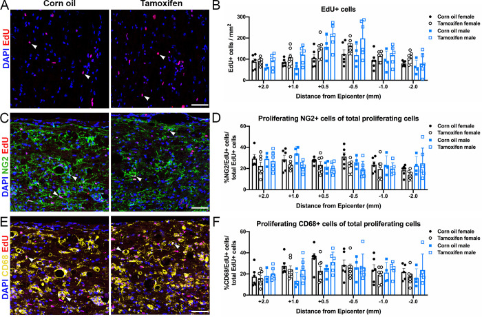
Fig 7. Delayed short-term tamoxifen treatment does not alter cell proliferation.
(A, C, E) Representative confocal images of (A) proliferating cells (EdU/DAPI+ cells), (C) proliferating NG2 cells (NG2/EdU/DAPI+ cells), and (E) proliferating phagocytic macrophages/monocytes (CD68/EdU/DAPI+ cells) in the lateral white matter of control and tamoxifen mice. Scale bar = 75 μm. (B) Quantification of the total number of proliferating cells 2 mm, 1mm, and 0.5 mm rostral and caudal to the lesion epicenter. (D, F) Percent of proliferating (D) NG2 cells and (F) phagocytic macrophages/monocytes normalized to total proliferating cells at each sample distance rostral and caudal to the lesion. (B, D, F) The data are not significantly different between control and tamoxifen mice or female and male mice at any distance from the lesion epicenter. Data presented as mean + SEM. Mixed-effects analysis with Šídák post hoc test; corn oil female n = 6; tamoxifen female n = 7; corn oil male n = 5; tamoxifen male n = 6.

