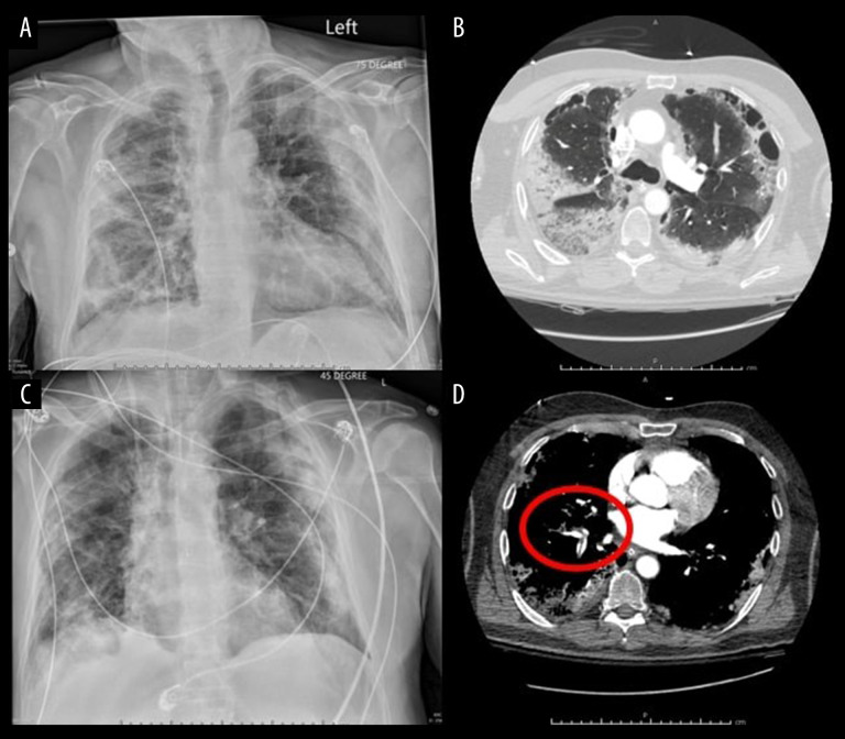
Figure 1.
Selected imaging studies from a 67-year-old man who presented to the hospital 2 weeks after testing positive for SARS-CoV-2. (A) Chest x-ray on presentation to the hospital; (C) Chest x-ray 5 days after admission to the hospital. (B, D) Computerized tomography angiography scan on presentation to the hospital showing diffuse lung disease (B) and pulmonary artery emboli (red circle) (D).

