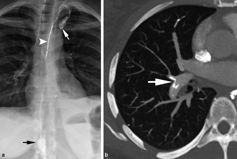
Fig. 7.

A 45-year-old woman with idiopathic chylothorax. ( a ) Following retrograde transvenous embolization of the thoracic duct with coils (white arrowhead) and glue, the glue migrated beyond the coil pack as the catheter was withdrawn, extending into the subclavian vein (white arrow). Black arrow—cisterna chyli. ( b ) The patient developed dyspnea 3 days postprocedure, and computed tomography of the chest was performed revealing multifocal glue segmental and subsegmental pulmonary emboli (white arrow).
