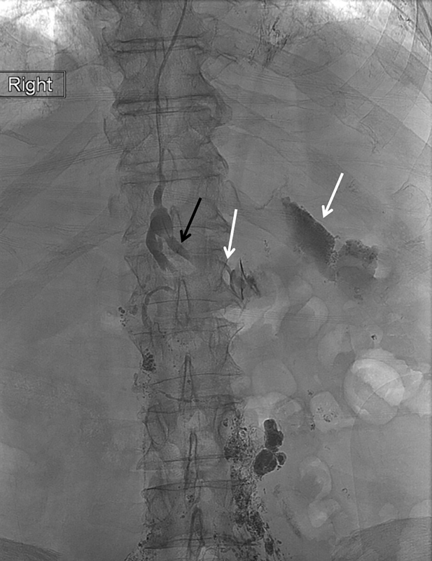
Fig. 19.

Spot radiograph of the upper abdomen during retrograde direct percutaneous lymphangiography with the tip of the microcatheter in the left lumbar trunk (black arrow) showing extravasation of contrast into the left upper quadrant (white arrows).
