
Fig. 1.
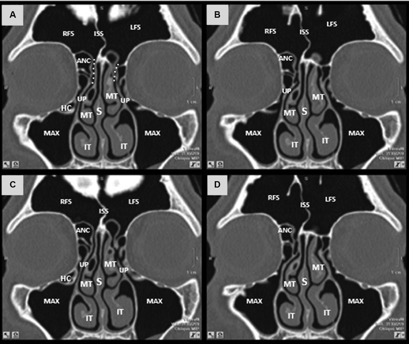
Coronal sections (A-D) computed tomography scan depicting the anatomy of the frontal sinus drainage pathway and surrounding structures. ANC = agger nasi cell; RFS = right frontal sinus; white dotted line = frontal sinus drainage pathway; ISS = intersinus septum; IT = inferior turbinate; LFS = left frontal sinus; Max = maxillary sinus; MT = middle turbinate; S = nasal septum; HC = haller cell; UP = uncinate process.