
Fig. 7.
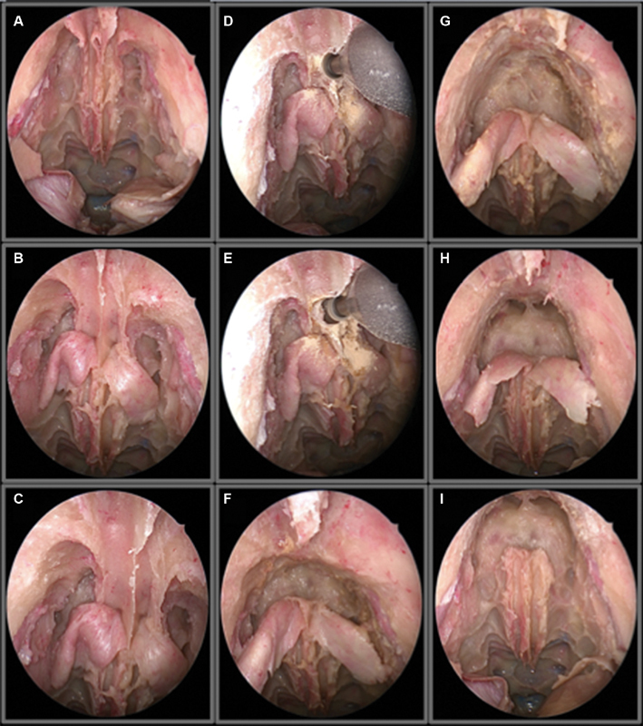
Endoscopic view of a Draf III as a step for a transcribriform approach. ( A ) view of the cribriform plate and frontal recess. ( B ) bilateral identification of the first olfactory fiber, ( C ) view after partial drilling of the frontal sinus floor, ( D ) drilling of the bony junction between the two frontal sinus, ( E ) visualization of the left frontal sinus, ( F ) view after communicating both frontal sinuses, ( G ) further drilling of the anterior table of the frontal sinus, ( H ) and ( I ) final aspect after a complete drill out of the frontal sinuses.