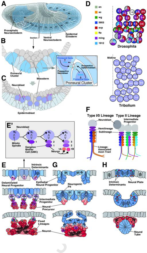
Figure 1.
Structure and origin of neural lineages in Drosophila. (A) Lateral surface view of Drosophila embryo at a stage shortly after gastrulation. Large ectodermal domain forming part of the head (procephalic neuroectoderm; origin of the brain) and the trunk (ventral neuroectoderm; origin of ventral nerve cord) gives rise to neural lineages. White lines and lettering delineate segmental units (An antennal segment; Ic intercalary segment; C2–3 gnathal segments; T1-T3 thoracic segments; A1–8 abdominal segments). (B, C) Cross sections of ventral neuroectoderm before (B) and after (C) neuroblast segregation. Expression of bHLH genes of the Achaete-Scute-Complex divide the neuroectoderm into an orthogonal pattern of proneural clusters (dark lilac). Notch-Delta signaling within proneural clusters (inset) selects one cell that delaminates as a neuroblast (C). (D) Top: Pattern of neuroblasts of one Drosophila abdominal hemisegment. Each neuroblast is a unique cell (1–1 to 7–4), defined by its time of delamination, position, and combinatorial expression of transcription factors and signaling molecules (see key to color code on the left of panel; after Doe, 1992, with permission). The neuroblast pattern is highly conserved among different insect taxa, as exemplified by the coleopteran Tribolium castaneum (bottom of panel; after Biffar and Stollewerk, 2014, with permission). (E, E’) Schematic cross-sections of insect embryo at sequential stages of development, illustrating characteristics of neural progenitors in insects. Top panel depicts epithelial neuroectoderm (blue) from which neural progenitors (lilac) originate. Insect neural progenitors (neuroblasts) delaminate and initiate a series of determinate, asymmetric divisions, giving rise to intermediate progenitors (magenta) called ganglion mother cells (GMCs). GMCs typically undergo one more mitosis into two neuronal (or glial) precursors (orange) which subsequently differentiate into neurons (red). Inset (E’) shows details of neuroblast division. Freshly delaminated neuroblast (N0) divides asymmetrically into apical second generation neuroblast (N1,2…) and basal ganglion mother cell (GMC1,2…). Ganglion mother cells undergo one terminal mitosis into molecularly different neurons (1A, 1B). (F) Structure of neural lineages in insects. Canonical proliferation pattern, delineated in panel (E), results in a type I lineage. Neurons born as “A” daughter cells and “B” daughter cells of ganglion mother cells represent the A and B hemilineages. Neurons born sequentially in a defined time window define a sublineage. Certain subsets of neuroblasts of the brain generate type II lineages, whereby neuroblasts produce intermediate progenitors, which, in turn, each behave like a type I neuroblasts, generating neurons via ganglion mother cells. (G, H) Generation and proliferation of neural progenitors in chelicerates (spiders; G) and urochordates (sea squirts; H). In these phyla, parts of the neuroectderm invaginate as multiple small “neurogenic pits” (in chelicerates, as well as other arthropod taxa) or single large neural tube (in urochordates, as well as the closely related vertebrates). Prior to the birth of neural precursors or intermediate progenitors, the neuroepithelium (blue) undergoes a phase of abundant symmetric divisions.

