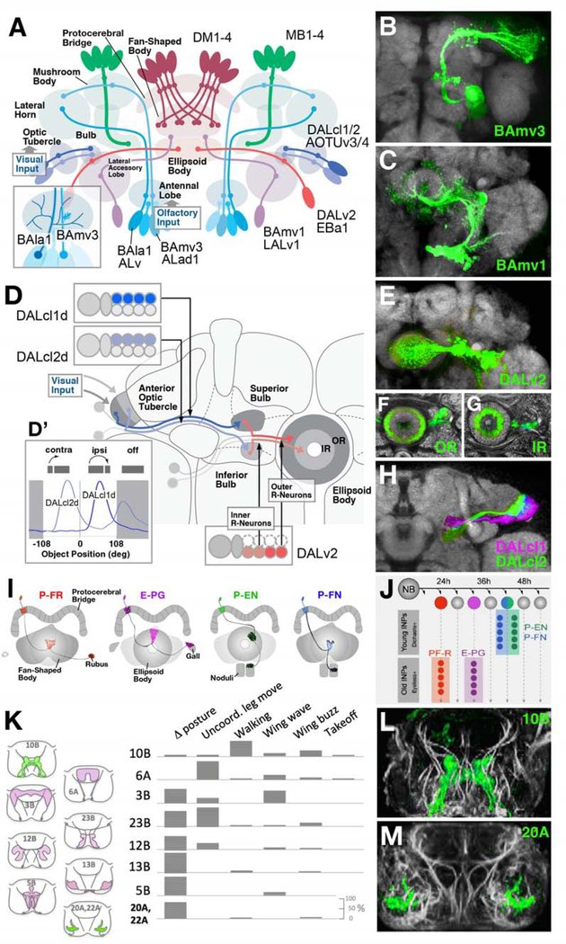
Figure 4.
Lineage-based architecture of the Drosophila brain. (A) Schematic of the Drosophila brain, illustrating representative lineages (BAla1/ALv, BAmv3/ALad1, BAmv1/LALv1, DALv2/EBa1, DALcl1/2/AOTUv3, MB1–4, DM1–4) and the compartments innervated by them. BAla1 and BAmv3 both form projection neurons connecting the antennal lobe (olfactory center) with the mushroom body; however, they differ in dendritic geometry (inset at lower left), with BAla1 forming wide-spread multiglomerular branches, and BAmv3 narrow, uni- or bi-glomerular branches. (B, C) Z-projections of frontal confocal sections of adult fly brain, illustrating GFP-labeled clones of lineages BAmv3/ALad1 and BAmv1/LALv1, respectively. (D-H) Lineage-based composition of the anterior visual pathway (AVP), which conducts visual information from the optic lobe via anterior optic tubercle and bulb to the ellipsoid body, as shown schematically in (D). Two hemilineages, DALcl1/AOTUv3 d and DALcl2/AOTUv4 d [inset at upper left of (D); GFP-labeled in confocal image shown in (H)] form parallel pathways between discrete subdomains of the tubercle and bulb. Based on Calcium-imaging of these neuron populations, DALcl1 d neurons react in a retinotopic manner to small stimuli in the ipsilateral visual field; DALcl2 d neurons do not show any retinotopy, and are active when stimulated contralaterally, as well as after cessation of the stimulus [inset at bottom left of (D)]. Lineage DALv2/EBa1 generates neurons that continue the parallel visual pathways from bulb to ellipsoid body (inset at bottom right of D). Early born (outer ring) neurons connect the superior bulb to the periphery of the ellipsoid body (OR); later born (inner ring) neurons project from inferior bulb to ellipsoid body center [IR; panels (F, G) and inset at bottom right of (D)]. (I, J) Sublineages of the type II neuroblasts DM1–4 form discrete classes of columnar neurons of the central complex (P-FR, E-PG, P-EN, P-FN), born during different time intervals from different intermediate progenitors (INPs; panel J). Early INP offspring are specified by expression of Dichaete; late offspring by Eyeless (from Sullivan et al., 2019; with permission). (K-M) Hemilineages form spatially and functionally discrete populations of interneurons in the ventral nerve cord. (K, Left) Schematic frontal sections of ventral nerve cord with domains innervated by lineages indicated (10B, 6A, 3B, 23B, 12B, 13B, 5B, 20A/22A) rendered in colors. (K, right) Types of behaviors preferentially elicited by stimulating hemilineage indicated. (L, M) Z-projections of frontal confocal sections of ventral nerve cord, showing GFP-labeled hemilineages 10B (elicits walking and wing beat) and 20A (involved in leg posture; from Harris et al., 2015, with permission).

