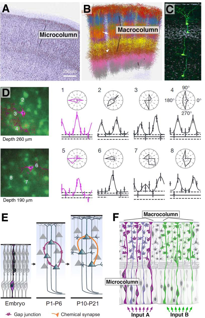
Figure 5.
Significance of cell lineage in the mammalian cerebral cortex. (A) Nissl-stained frontal section of the human cerebral cortex, showing columnar arrangement of neuronal cell bodies (from Jones, 2000, with permission). (B) Digital 3D reconstruction of five neighboring barrel columns in rat somatosensory cortex. Branched neurite trees of different types of neurons are rendered in different colors (after Egger et al., 2014; with permission by Dr. Marcel Oberlaender). Macrocolumns are spatially well separated in cortical layer IV (arrow), but not in deep or superficial layers (arrowhead). (C) Neurons derived from one apical radial glia progenitor at the onset of neurogenic divisions form a coherent ontogenetic column (GFP-labeled; from Gao et al., 2014, with permission). (D) Sibling neurons forming part of one ontogenetic column have direction preference. Shown at the left are tangential confocal sections of the visual cortex at two different depths. Sibling neurons appear in magenta, general neurons in green. To the right are polar plots of orientation tuning of neurons #1–8. Note similar tuning of siblings #1 and 5 (from Li et al., 2012, with permission). (E) Sibling neurons are strongly electrically coupled by gap junctions (purple) during the first postnatal weeks (P1-P6). At a later stage, the same neurons form preferentially chemical synapses (orange) among themselves (from Gao et al., 2013, with permission). (F) Schematic representation of relationship between microcolumn and macrocolumn. Numerous adjacent microcolumns, representing developmentally based ontogenetic columns, are bundled into larger units (macrocolumns) by shared thalamic input (e.g., afferents from a single vibrissa) or other connections.

