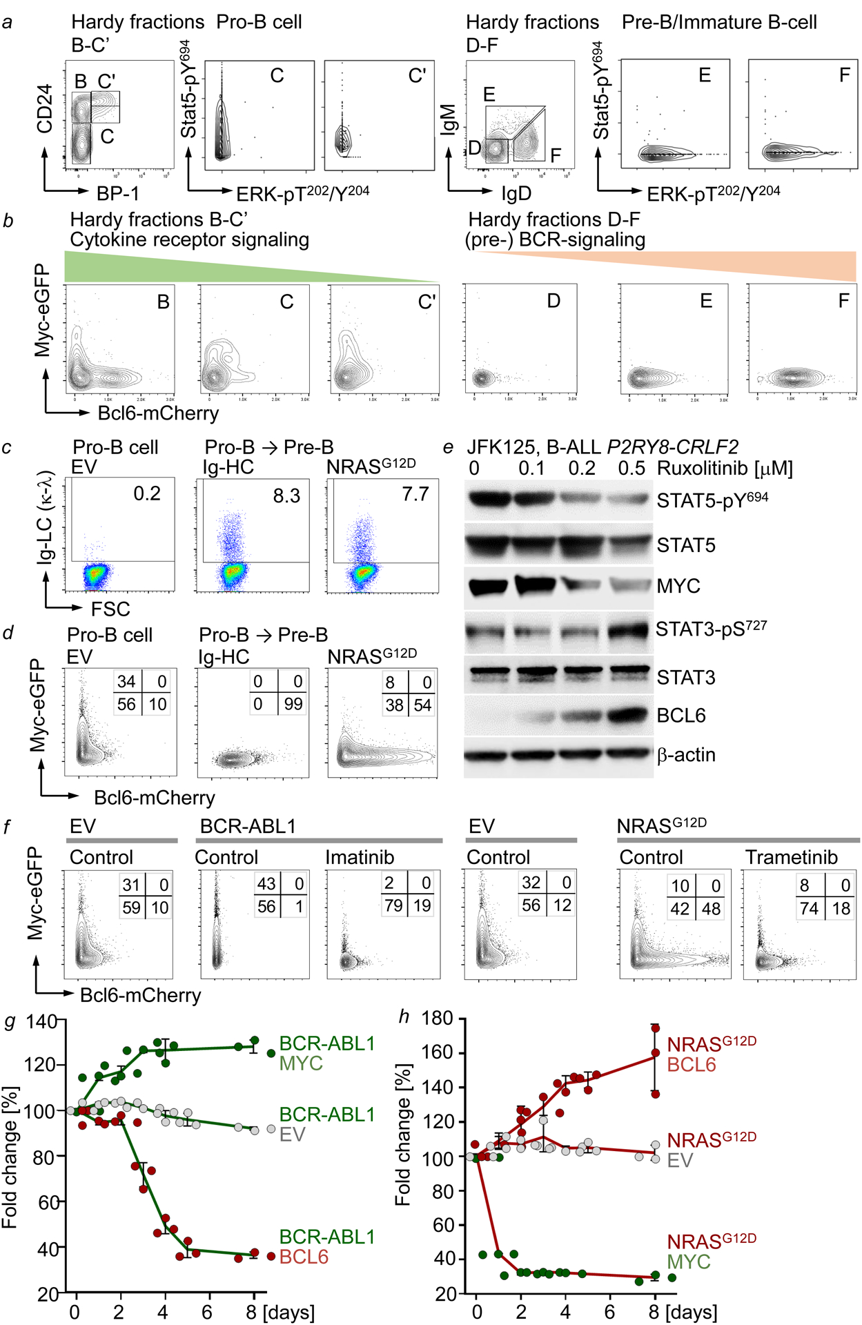
Figure 2: STAT5-MYC and ERK-BCL6 signaling are incompatible and define distinct stages of B cell development.

a, Hardy Fractions B-F analysis of MyceGFP/+ Bcl6mCherry/+ bone marrow cells. Single-cell phosphoprotein analyses of the indicated fractions. b, Surface expression of eGFP and mCherry on MyceGFP/+ Bcl6mCherry/+ bone marrow cells in Hardy Fractions B-F. c, Surface expression of Igκλ light chain (LC) on IL7-dependent pro-B cells (left), and upon Dox-inducible expression of μHC (middle) or NRASG12D (right). d, Surface expression of eGFP and mCherry on IL7-dependent MyceGFP/+ Bcl6mCherry/+ B-cell precursors induced to differentiate or transduced with NRASG12D. e, Western blots of Ph-like B-ALL cells treated with ruxolitinib (n=3 independent experiments). f, Surface expression of eGFP and mCherry on MyceGFP/+ Bcl6mCherry/+ B-cell precursors transduced with EV, BCR-ABL1 or NRASG12D, and subsequently treated with vehicle control, imatinib (1 μM) or trametinib (10 nM). g,h, Enrichment or depletion of GFP+ BCR-ABL1 or NRASG12D B-ALL cells transduced with EV, GFP-MYC or GFP-BCL6 (mean, ±s.d; g,h). Data from 3 independent biological replicates (a-d,f). e-h, n=3 independent experiments. For gel source data, see Supplementary Fig. 1.
