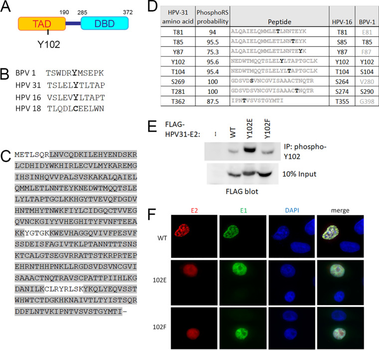
FIG 1.
Identification of phosphorylated Y102. (A) Schematic of HPV-31 E2 protein representing the transactivation domain (TAD) and DNA-binding domain (DBD). (B) Conservation of the region surrounding Y102. (C) HPV-31 E2 was purified, digested with trypsin, and submitted for mass spectrometric analysis. Peptide coverage was 95% as highlighted in gray. (D) Phosphorylated amino acids of HPV-31 E2 with high PhosphoRS probabilities (above 75%) are listed on the left. The trypsin fragments carrying the modifications are listed in the middle, with phosphorylation sites in black. HPV-16 and BPV-1 homologue residues are listed on the right, with conserved residues in black and nonconserved in gray. (E) FLAG-HPV-31 E2 (WT or Y102 mutants) was expressed in HEK 293TT cells, immunoprecipitated with custom phospho-specific Y102 antibody, and blotted with FLAG antibody. (F) Y102 mutants localize to the nucleus. N/TERT cells were transfected with FLAG-HPV-31 E2 (WT, Y102E, and Y102F) and HA-HPV-31 E1, and immunostained with FLAG and HA antibodies.

