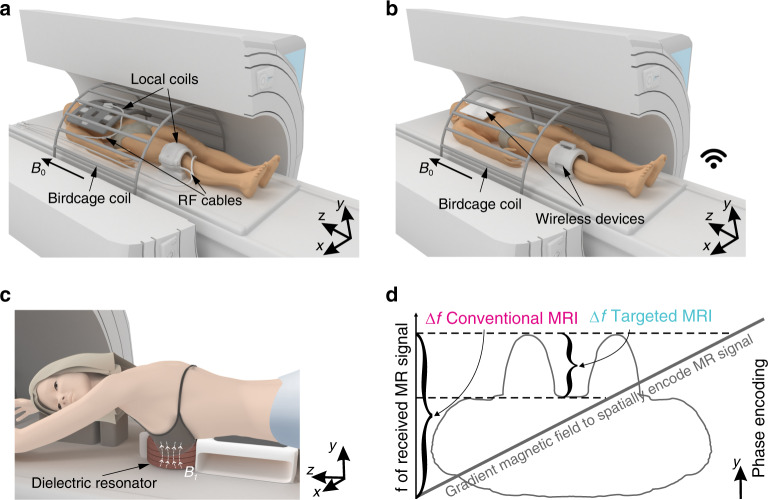
Fig. 1. Conventional and targeted concepts of MRI.
a An illustration of a conventional acquisition scheme in the clinical MR systems. A body-sized RF resonator (“birdcage coil”) placed in the wall of an MR system bore excites the MR signal, while multiple surface RF coils located directly on a patient (“local coils”) receive MR signal and cabling system through a patient table delivers it to a spectrometer. B0 indicates the direction of the static magnetic field of an MR system. b A targeted MRI concept. The same large body resonator excites and receives MR signal while a wireless device localizes both exciting and receiving RF magnetic fluxes of the body resonator to the region of interest. c An illustration of the actual experimental setup of targeted MRI for breast at 3 Tesla: the RF magnetic field (B1-field) is confounded to the dielectric resonator cavity where a breast is located. d The gradient magnetic fields are always used during MR examinations to encode the MR signal through its frequency and phase. In the targeted MRI, the received signal bandwidth is limited to the size of the area of interest, while in the conventional MRI, the signal bandwidth is always proportional to patient body size. A significant reduction in the received signal bandwidth (in the phase-encoding direction) is beneficial for the total acquisition time of high-resolution imaging of the area of interest because of a decreased Nyquist frequency in the phase encoding direction.

