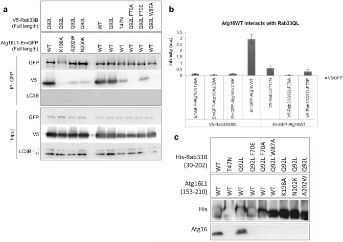
Figure 5.
Effects of Rab33B and Atg16 mutations on complex formation analyzed by GFP co-immunoprecipitation and Ni-sepharose pulldown experiments. (a) GFP Co-immunoprecipitation. Overexpression was done in HEK293 cells. Western blots were probed with either anti-GFP, anti-V5 or anti-LC3B antibodies. IP immunoprecipitation. (b) Quantification of the western blot band intensities shown in (a). Band intensities were calculated by normalizing the V5-IP blot band intensities against GFP-IP intensity. Error bars represent standard errors (SE) of three independent biological replicates. (c) His-Rab33B(30–202) mutants were co-expressed with Atg16L1(153–210) wild-type and mutants. Samples were run on Schägger gels after elution from the Ni-Sepharose beads and then blotted onto nitrocellulose membranes. Membranes were probed with rabbit anti-Atg16L primary antibody and goat anti-rabbit IgG (HRP labeled) secondary antibody. Uncropped images of blots are shown in Figures S7 and S8.

