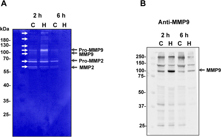
Figure 5.
Activation of mouse skin proteinases. (A) Gelatin zymography of mouse skin proteins. Protein samples (30 μg) from control skin proteins (C) or hemorrhagic skin proteins (H), prepared under non-reducing conditions, were submitted to electrophoresis on 10% SDS–polyacrylamide copolymerized with gelatin. Gels were stained with Coomassie blue. Proteins with activity were identified as clear zones of lysis against a dark background. White arrows indicate bands of gelatinolytic activity. (B) Detection of the presence of MMP9 in the mouse skin, as shown by Western blot analysis. Protein samples (30 μg) from control skin proteins (C) or hemorrhagic skin proteins (H) were immunostained with anti-MMP9 antibodies. Numbers on the left indicate molecular mass marker mobility.

