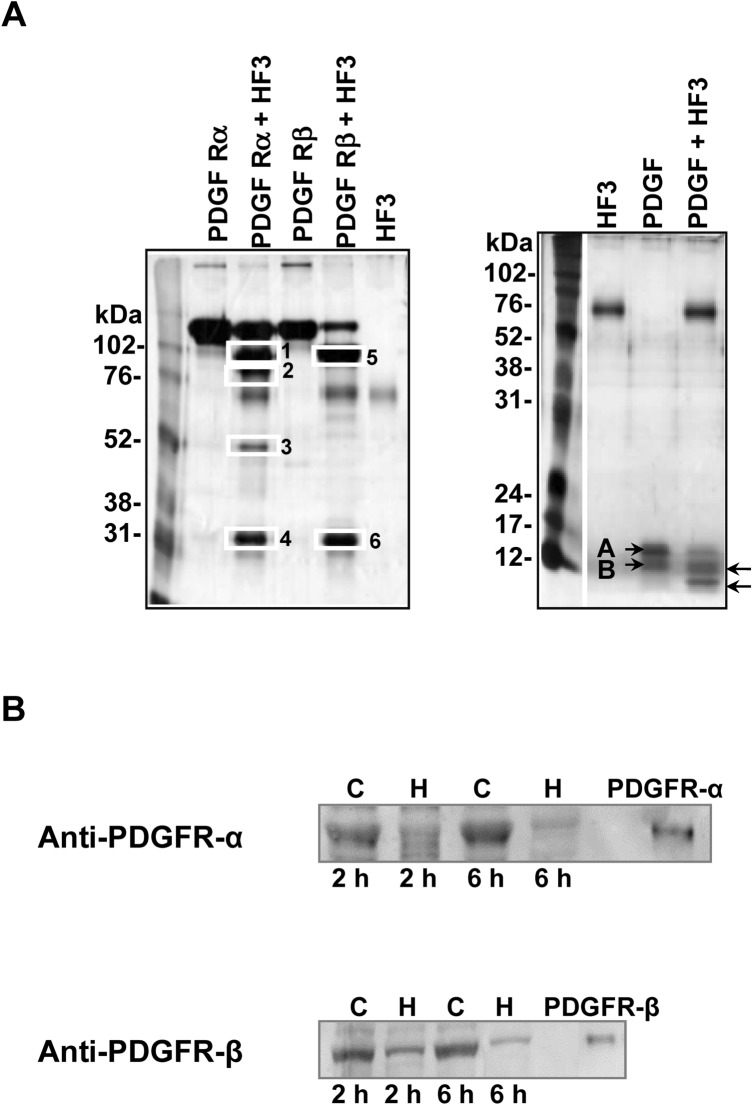
Figure 7.
Activity of HF3 upon PDGFR and PDGF. (A) HF3 cleaves PDGFR (alpha and beta forms) and PDGF in vitro. PDGFR-α (recombinant mouse PDGFR-α-Fc chimera), PDGFR-β (recombinant mouse PDGFR-β-Fc chimera) and PDGF were incubated with HF3, as described in “Materials and methods”, and submitted SDS-PAGE. White rectangles indicate proteins bands identified by mass spectrometry. Letters A and B indicate the subunits of the disulfidelinked PDGF heterodimer. Arrows indicate cleavage products generated by HF3. Proteins were stained with silver. (B) Degradation of PDGFR-α and PDGFR-β in the mouse skin injected with HF3, as shown by Western blot analysis. Protein samples (30 μg) from control skin (C) or skin injected with HF3 (H), and PDGFR-α (recombinant mouse PDGFR-α-Fc chimera) and PDGFR-β (recombinant mouse PDGFR-β-Fc chimera) (10 ng) were immunostained with anti-PDGFR-α and anti-PDGFR-β antibodies. Full-length images of Western blots and SDS-PAGE profiles of mouse proteins from control and hemorrhagic skins in comparison with PDGFRα and PDGFRβ are shown in Supplementary Figure S3.

