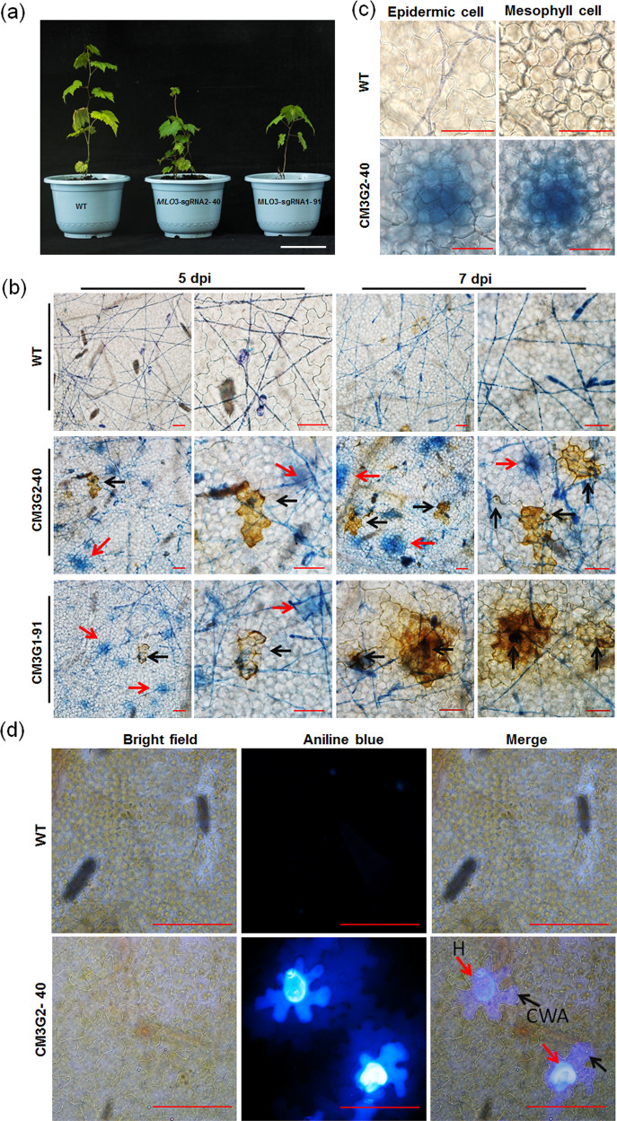
Fig. 6. VvMLO3-edited grapevine plants show infection-triggered cell death, H2O2 accumulation and CWAs.
a The wild-type (WT) and two VvMLO3-edited heterozygous mutant grapevine lines grown under phytotron conditions for 6 months (bar = 10 cm). b Representative micrographs showing DAB- and trypan blue-stained epidermal cells of the WT and VvMLO3-edited lines at 5 or 7 dpi. Red arrowheads indicate trypan blue retention, and black arrowheads indicate H2O2 accumulation (bar = 50 μm). c Representative images showing a trypan blue-stained leaf section of WT or CM3G2-40 with a focus on either the epidermal layer or the mesophyll cell layer at 7 dpi (bars = 50 μm). d Histochemical analysis of infection-triggered CWAs of epidermal cells of the WT and the heterozygous CM3G2-40 mutant line at 7 dpi. Red arrowheads indicate haustoria (H), and black arrowheads indicate infection-triggered CWAs (bars = 50 μm).

