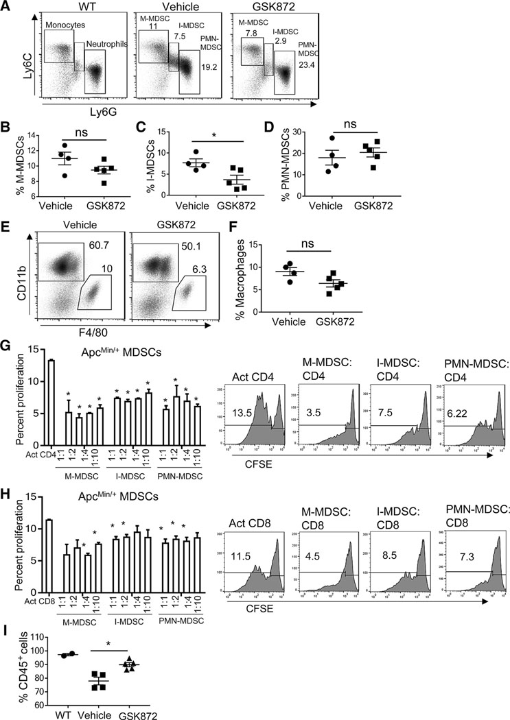
Figure 2.
I-MDSCs are reduced in ApcMin/+ mice treated with GSK 872. A, Representative flow cytometry plots of WT monocytes and neutrophils, and ApcMin/+ MDSC subsets; percentages of M-MDSCs (B), I-MDSCs (C), and PMN-MDSCs (D) gated on CD45+CD11bhiCD11clo live splenocytes, macrophages (CD11b+F4/80+; E and F), and CD45+ (I) cells from ApcMin/+ mice receiving vehicle control (n = 4) or GSK 872 (n = 5). G and H, ApcMin/+ MDSC subsets suppress CFSE-labeled, polyclonally activated (anti-CD3 and anti-CD28 antibodies) WT, CD4, and CD8 T-cell proliferation. Representative flow cytometry plots show the suppression of CD4 and CD8 T cells by MDSCs (1:1 MDSCs to T cells). Activated T cells without MDSC coculture were positive controls. Comparisons were made between positive control and each MDSC to T-cell coculture. *, P < 0.05; ns, nonsignificant.

